


Most bladder cancers arise from the cells that form the innermost layer of the bladder. These cells, called transitional cells or urothelial cells, allow the bladder to stretch when it is full and shrink when it is emptied. Transitional cells are also the type of cells responsible for most cancers of the renal pelvis and ureters.
Bladder cancer most often causes blood in the urine.
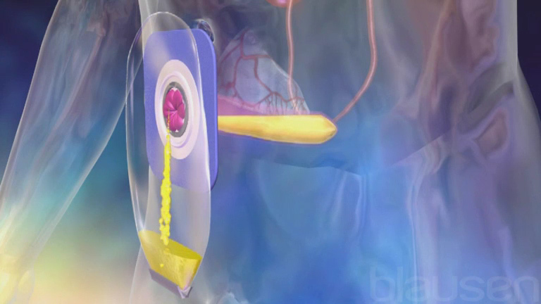
To make the diagnosis, doctors insert a thin, flexible tube with a camera (cystoscope) through the urethra into the bladder.
Treatment often involves removing the cancer, using a cystoscope (for surface cancers) or surgically removing the entire bladder (for deeper cancers).
About 82,290 new cases of bladder cancer are diagnosed every year in the United States. According to 2023 estimates, more than 16,710 people die of bladder cancer every year. About 3 times as many men as women develop bladder cancer.
The chronic irritation that occurs with a parasitic infection called schistosomiasis or with bladder stones, urinary tract infections, or chronic catheter use also predisposes people to bladder cancer, although irritation accounts for only a small number of all cases.

Symptoms of Bladder Cancer
Bladder cancer most often causes blood in the urine. Other symptoms may include pain and burning during urination and an urgent, frequent need to urinate. The symptoms of bladder cancer may be identical to those of a bladder infection (cystitis), and the problems may occur together. A low blood count (anemia) may cause fatigue, paleness, or both.
Diagnosis of Bladder Cancer
Blood in urine
Cytology (examination of urine under the microscope)
Cystoscopy (visualization of the inside of the bladder) and biopsy (examination of bladder tissue under the microscope)
The diagnosis is often first suspected when blood is found in the urine. Blood may be detected when a routine microscopic examination of a urine specimen detects red blood cells, or sometimes the urine may be visibly red. Bladder cancer may be suspected if the symptoms of cystitis do not disappear with treatment. Special microscopic evaluation of urine (such as cytology) may detect cancer cells. Sometimes bladder cancer is detected when an imaging study such as computed tomography (CT) or ultrasonography is done for another reason.
Most bladder cancers are diagnosed by cystoscopy and biopsy. This examination involves passing a thin, flexible tube with a camera (cystoscope) through the urethra into the bladder. If anything is abnormal, a biopsy may be done in the operating room under anesthesia using a special cystoscope.
If the cancer has invaded the bladder muscle, additional testing, including abdominal CT and chest x-ray, is needed to determine whether a cancer has spread. Magnetic resonance imaging (MRI) can now be used to determine the extent of spread in the area around the bladder cancer.
Improvements in detecting and staging bladder cancer are expected to improve outcomes by finding the cancer earlier.
Treatment of Bladder Cancer
Removal during cystoscopy
Intravesical immunotherapy or chemotherapy (for superficial, or surface, cancers)
Partial or total removal of the bladder, radiation, chemotherapy or immunotherapy (for deeper, more invasive cancers)
Cancers that have grown into the bladder wall cannot be completely removed through a cystoscope. They are usually treated by total or partial removal of the bladder (cystectomy). Chemotherapy is usually given before removing the bladder as this has been shown to improve survival compared to cystectomy alone. Radiation therapy alone or in combination with chemotherapy is used in an attempt to cure the cancer in selected people.
If the entire bladder needs to be removed, doctors must devise a method for the person to be able to drain urine. The usual way has been to route the urine to an opening (stoma) made in the abdominal wall through a passageway made of intestine, called an ileal loop (conduit). The urine is then collected in a bag worn outside the body.
Several alternative methods of diverting urine are becoming increasingly common and are appropriate for many people. These methods can be grouped into 2 categories: an orthotopic neobladder and a continent urinary diversion. In both, an internal reservoir for urine is constructed from the intestine.
For an orthotopic neobladder, the reservoir is connected to the urethra. The person learns to empty this reservoir by relaxing the pelvic floor muscles and increasing pressure within the abdomen, so that urine passes through the urethra very much as it would naturally. Most people are dry during the day, but some urine leakage may occur at night.
For a continent urinary diversion, the reservoir is connected to a stoma in the abdominal wall. A collecting bag is not needed, because the urine remains in the reservoir until the person empties it by inserting a catheter through the stoma into the reservoir, which is done at regular intervals throughout the day. The most common of these is known as an Indiana pouch and is made from a part of the colon.
Cancer that has spread beyond the bladder to the lymph nodes or other organs is treated with chemotherapy. Several different combinations of medications are active against this type of cancer, particularly when the spread is confined to the lymph nodes. Cystectomy or radiation therapy, including external beam radiation, may be offered to people who respond well to chemotherapy. However, a relatively small number of people are cured. For people who are not cured, efforts are directed at pain relief (see Symptoms During a Fatal Illness) and end-of-life issues.
Prognosis for Bladder Cancer
For cancers that remain on the bladder’s inner surface (superficial tumors) and grow and divide slowly, the risk of death from bladder cancer is less than 5% in the 5 years after diagnosis. The 5-year death rate for tumors that invade the bladder muscle is significantly higher (about 50%), but chemotherapy may improve survival. Cancers that have spread beyond the bladder wall (such as to the lymph nodes or other abdominal or pelvic organs) have a much poorer prognosis.


