


Atelectasis is a condition in which all or part of a lung becomes airless and collapses.
Blockage of the bronchial tubes is a common cause of atelectasis.
Shortness of breath can develop if oxygen levels are low or pneumonia occurs.
Chest x-ray is used to confirm the diagnosis.
Treatment may involve making sure deep breathing occurs, relieving airway blockages, or both.
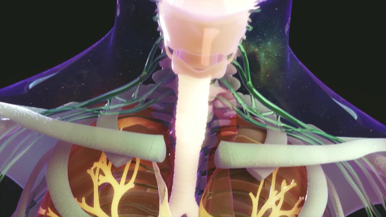
The main function of the lungs is to absorb oxygen into the bloodstream from the atmosphere and to expel carbon dioxide from the blood into the exhaled breath (gas exchange—see figure Gas Exchange Between Alveolar Spaces and Capillaries). For gas exchange to occur, the small air sacs within the lungs (alveoli) must remain open and filled with air. Alveoli are kept open by the elastic structure of the lung and by a liquid lining called surfactant. Surfactant counters the natural tendency of the alveoli to close (collapse). Periodic deep breaths, which people take unconsciously, and coughing also help keep alveoli open. Coughing expels any mucus or other secretions that could block the airways leading to the alveoli.
If the alveoli are closed for any reason, they cannot participate in gas exchange. The more alveoli that are closed, the less gas exchange occurs. Accordingly, atelectasis can decrease the level of oxygen in the blood. The body compensates for a small amount of atelectasis by closing off (constricting) the blood vessels in the affected area. This constriction redirects blood flow to alveoli that are open so that gas exchange can continue.
Causes of Atelectasis
Common causes of atelectasis usually involve one of the following
Blockage of one of the tubes (bronchi) that branch off from the trachea (windpipe) and lead to the lung tissue
Conditions that decrease deep breathing or suppress a person’s ability to cough
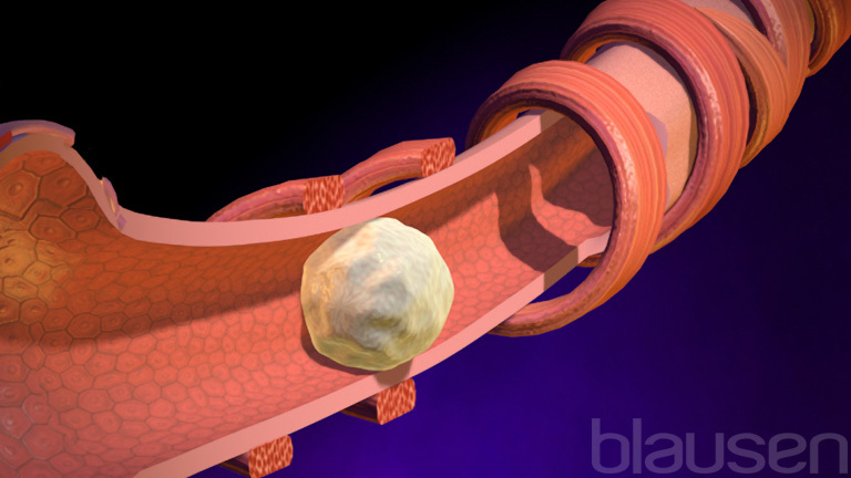
The blockage may be caused by something inside the bronchus, such as a plug of mucus, a tumor, or an inhaled foreign object (such as a pill, a piece of food, or a toy). Alternatively, the bronchus may be blocked by something pressing from the outside, such as a tumor or an enlarged lymph node. Blockage from the outside can also occur if the pleural space (the space outside of the lung but inside of the chest) contains a large amount of fluid (pleural effusion) or air (pneumothorax).
When a bronchus or a smaller airway (bronchiole) becomes blocked, the air in the alveoli beyond the blockage is absorbed into the bloodstream, causing the alveoli to shrink and collapse. The area of collapsed lung may become infected because bacteria and white blood cells can build up behind (to the inside of) the blockage. Infection is particularly likely if atelectasis persists for several days or more. If atelectasis persists for months, the lung may not easily re-expand.
Any condition that decreases deep breathing or suppresses a person’s ability to cough can cause or contribute to atelectasis. Large doses of opioids or sedatives can decrease deep breathing. Atelectasis is common after general anesthesia, which temporarily suppresses a person’s cough and drive to breathe. Atelectasis is particularly common after chest or abdominal surgery because the effects of receiving general anesthesia may be added to the pain of deep breathing, so people take only shallow breaths. Chest or abdominal pain due to other causes (for example, injury or pneumonia) also makes taking a deep breath painful.
Certain neurologic conditions, immobility, and chest deformities can limit chest movement and thus decrease deep breathing, as can abdominal swelling. People who are very overweight or obese are also at greater risk of developing atelectasis.
Did You Know...
|
Symptoms of Atelectasis
Atelectasis itself does not cause any symptoms except sometimes shortness of breath. The presence and severity of shortness of breath depend on how rapidly atelectasis develops and how much of the lung is affected. If atelectasis involves a limited portion of the lung or develops slowly, symptoms may be mild or not even noticed. If a large number of alveoli are affected, particularly if atelectasis occurs rapidly, shortness of breath may be severe.
The heart rate and breathing rate may increase, and sometimes the person may look bluish (a condition called cyanosis) because oxygen levels in the blood are low. This appearance may be darker brown or black in people with dark skin.
Symptoms may also reflect the disorder that caused atelectasis (for example, chest pain due to an injury) or a disorder that results from atelectasis (for example, chest pain with deep breathing, due to pneumonia).
Diagnosis of Atelectasis
Chest x-ray
Doctors suspect atelectasis based on a person’s symptoms, the physical examination findings, and the setting (for example, after surgery, chest injury, or use of certain drugs) in which the symptoms occurred. A chest x-ray that shows the airless area confirms the diagnosis. Sometimes computed tomography (CT), bronchoscopy (inserting a viewing tube into the bronchus), or both may be done to find the cause.
Treatment of Atelectasis
Deep breathing and coughing
Relief of airway blockages by suctioning or bronchoscopy
Treatment of atelectasis may involve making sure deep breathing occurs, relieving airway blockages, or both.
Sometimes blockages can be relieved when a patient’s airway is suctioned by a health care practitioner. A blockage that cannot be removed by suctioning may require removal by bronchoscopy. Sometimes other methods are necessary. For example, if a tumor is blocking an airway, the blockage can sometimes be relieved by surgery, radiation therapy, chemotherapy, or laser treatment. If mucus is plugging the airways, doctors sometimes give drugs to try to thin the mucus or open the airways.
Treatment of complications
Symptoms and complications of atelectasis may require treatment. People may require
Supplemental oxygen
Antibiotics, if bacterial infection is suspected
Rarely, insertion of a breathing tube (endotracheal intubation) and mechanical ventilation
Prevention of Atelectasis
Atelectasis may be prevented by making sure deep breathing occurs. Whenever possible, conditions that cause shallow breathing for long periods should be treated.
People who smoke can decrease their risk of atelectasis after surgery by stopping smoking, ideally 6 to 8 weeks before surgery. All people who have surgery should be encouraged to breathe deeply, cough regularly, and move about as soon as possible after the operation. The use of devices to encourage voluntary deep breathing, called incentive spirometry, and certain exercises, including changing position to increase the drainage of lung mucus and other secretions, may also help prevent atelectasis.
More Information
The following English-language resource may be useful. Please note that THE MANUAL is not responsible for the content of this resource.
National Heart. Lung, and Blood Institute: Atelectasis: Discussion of diagnosis, including screening, as well as prevention and treatment


