


Injuries and disorders can put pressure on the spinal cord, causing back or neck pain, tingling, muscle weakness, and other symptoms.
The spinal cord may be compressed by bone, blood (hematomas), pus (abscesses), tumors (cancerous or not), or a ruptured or herniated disk.
Symptoms, such as back or neck pain, abnormal sensations, muscle weakness, or impaired bladder and bowel control, may be mild or severe.
Doctors base the diagnosis on symptoms and the results of a physical examination, magnetic resonance imaging, or other imaging tests.
Corticosteroids are often given to reduce swelling in or around the spinal cord and thus help reduce pressure on the spinal cord.
Depending on the cause, surgery and/or radiation therapy may be used to relieve the pressure.
(See also Overview of Spinal Cord Disorders.)
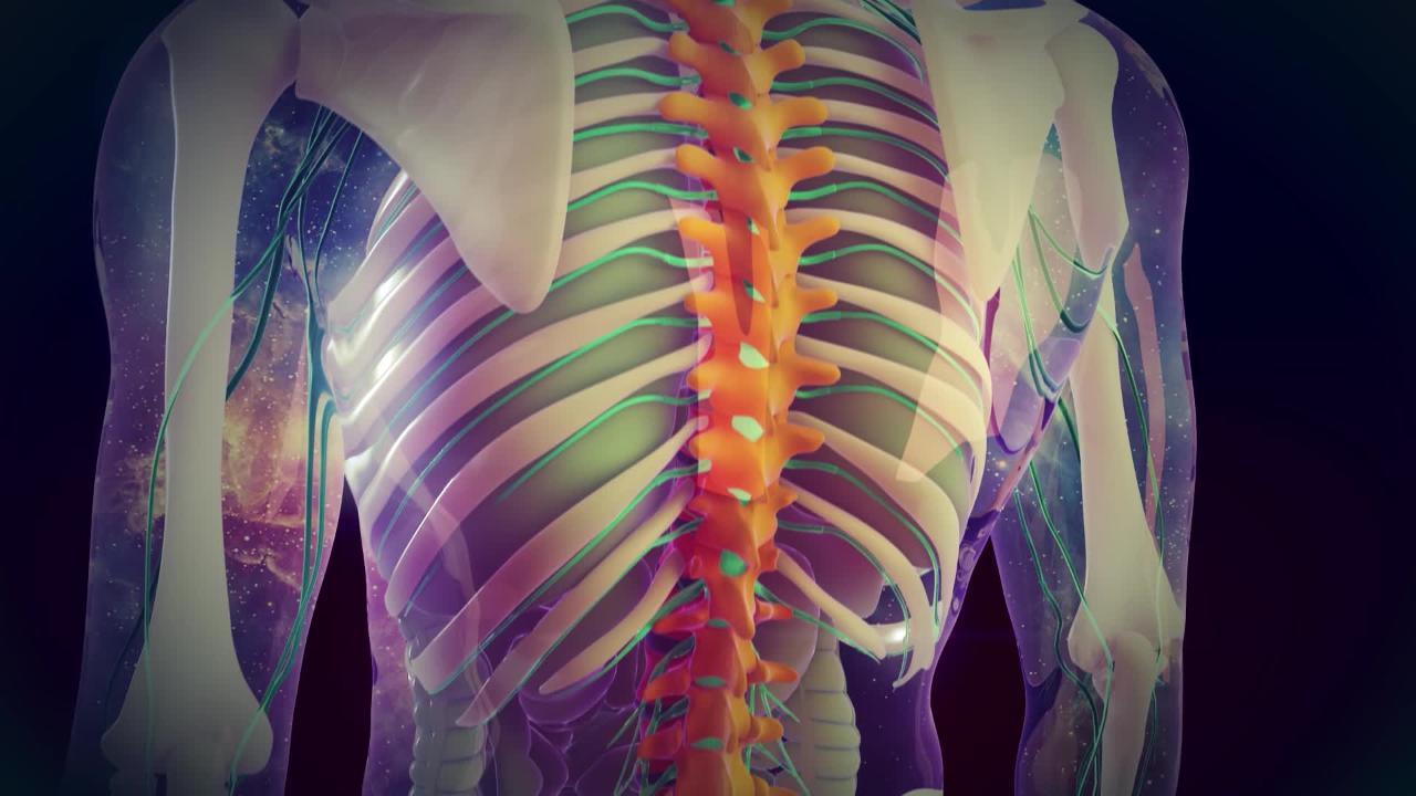
Normally, the spinal cord is protected by the spine, but certain injuries and disorders may put pressure (compress) on the spinal cord, disrupting its normal function. These injuries and disorders may also compress the roots of spinal nerves, which pass through the spaces between the back bones (vertebrae), or the bundle of nerves that extend downward from the spinal cord (cauda equina).
Spinal cord compression may occur
Suddenly, causing symptoms in minutes or over a few hours or days to weeks
Slowly and gradually, causing symptoms that worsen over months to years
Causes of Spinal Cord Compression
The spinal cord may be compressed by the following:
Bone: If the vertebrae are broken (fractured), are dislocated, or grow abnormally (as occurs in cervical spondylosis), they may compress the spinal cord. Vertebrae that are weakened by cancer or osteoporosis may break after a slight or even no injury.
Connective tissue: Connective tissue that lines the spinal canal often enlarges and hardens as people age. This change narrows the spinal canal and compresses the spinal cord. (The spinal canal is the passageway that runs through the center of the spine and contains the spinal cord.)
An accumulation of blood (hematoma): Blood may accumulate in or around the spinal cord. The most common cause of a spinal hematoma is an injury, but many other conditions can cause hematomas. They include abnormal connections between blood vessels (arteriovenous malformations), tumors, bleeding disorders, and use of anticoagulants (which interfere with blood clotting) or thrombolytic medications (which break up blood clots).
Tumors: Cancer that has spread (metastasized) to the spine or the space around the spinal cord is a common cause of compression. Rarely, a tumor within the spinal cord causes compression. The tumor may be cancerous or not.
A pocket of pus (abscess): Pus may accumulate outside the spinal cord or, less commonly, in the spinal cord and may compress it.
A ruptured or herniated disk: A herniated disk can compress spinal nerve roots (the part of spinal nerves next to the spinal cord) and occasionally the spinal cord itself.
How quickly compression occurs varies.
Compression that develops in minutes or a few hours typically results from
An injury (the most common cause), which often results in a fracture or dislocation of a vertebra
Hematomas
Ruptured disks
However, bones weakened gradually (for example, by cancer or osteoporosis) may suddenly fracture, which can suddenly cause or worsen compression (see Compression Fractures of the Spine).
Compression that develops over days to weeks typically results from
Metastatic tumors
Abscesses
At some point, compression by a tumor or abscess starts to block blood flow to the spinal cord. Lack of blood injures the spinal cord and causes it to swell. The swelling blocks blood flow even more, creating a vicious circle of more and more swelling of and injury to the cord. This vicious circle may develop over a few days, hours, or sometimes minutes. It is a major medical emergency that requires immediate treatment.
Compression may develop gradually, over months to years. Typical causes include
Cervical spondylosis (degeneration of vertebrae and disks in the neck)
Some slow-growing tumors
Symptoms of Spinal Cord Compression
Slight compression may cause mild symptoms if it disrupts only some nerve impulses going up and down the spinal cord. These symptoms may include
Discomfort or pain in the back or neck
Slight muscle weakness
Tingling or other changes in sensation
Other changes in sensation
In men, difficulty initiating and maintaining an erection (erectile dysfunction)
If the spinal cord in the low back is compressed, pain may radiate down a leg, sometimes to the foot. If the spinal cord in the neck is affected, pain may radiate down the arms. If the cause is cancer, an abscess, or a hematoma, the back or neck may be tender to the touch in the affected area. Sometimes sensation is lost. Reflexes, including the urge to urinate, may be lost. If compression increases, symptoms may worsen.
Substantial compression may block most nerve impulses, causing
Severe muscle weakness
Numbness
Retention of urine
Loss of bladder and bowel control
If all nerve impulses are blocked, the following result:
Paralysis (which can cause problems with breathing if the spinal cord in the neck is compressed)
Complete loss of sensation in the area controlled by the part of the spinal cord below the compressed part
Once compression begins to cause symptoms, the damage can worsen, depending on the cause, within minutes or over hours to days.
Diagnosis of Spinal Cord Compression
Physical examination
Magnetic resonance imaging or myelography with computed tomography
People with symptoms suggesting spinal cord compression require immediate medical attention because prompt diagnosis and treatment may reverse or lessen loss of function.
Because the spinal cord is organized in a specific way, doctors can determine which part of the spinal cord is affected based on the symptoms and results of a physical examination. For example, if the legs (but not the arms) are weak and numb and bladder and bowel functions are impaired, the spine may be damaged at the midchest (thoracic) or lower back (lumbar) level. The location of pain or tenderness along the spine also helps doctors determine the site of the damage.
Magnetic resonance imaging (MRI) is done immediately if possible. Or if MRI is unavailable, myelography with computed tomography (CT) is done. These tests usually show where the spinal cord is compressed and may indicate the cause. These tests can detect a fracture or dislocation of a vertebra, a herniated disk, an abnormal bone growth, an area of bleeding, an abscess, or a tumor. Myelography with CT involves doing CT of the spinal column after a spinal tap (lumbar puncture) is done to inject a small amount of radiopaque contrast agent, which can be seen on x-rays, into the space around the cord. Thus, doctors can determine whether compression completely blocks the normal flow of cerebrospinal fluid through this space.
If the cause is thought to be a fracture or dislocation due to injury, x-rays may also be taken. They provide information quickly, enabling doctors to quickly evaluate the problem.
The cause of the compression can be confirmed during surgery to relieve pressure on the spinal cord.
If MRI or myelography with CT detects an unidentifiable abnormal mass causing compression, doctors first decide whether it needs to be removed. If not, doctors usually do a biopsy. They may remove a sample of tissue for testing by inserting a needle into the mass (usually guided by CT) or sometimes by doing a surgical procedure.
Treatment of Spinal Cord Compression
Usually surgery
Sometimes corticosteroids given intravenously
For tumors, usually radiation therapy (with or without surgery)
For abscesses or hematomas, sometimes drainage
If loss of function is partial or very recent (usually when compression occurs suddenly), the compression must be relieved immediately. When compression is detected and treated quickly, before nerve pathways are destroyed, treatment can prevent permanent damage to the spinal cord, and function is usually recovered. Surgery is typically needed to relieve compression. Surgery may also be needed to insert rods, screws, and/or pins and thus stabilize the spine.
Other treatment varies depending on the cause.
Surgery is done in the following cases:
Symptoms worsen despite treatment.
A biopsy is needed.
The spine is unstable.
Tumors are present or recur after radiation therapy.
Doctors suspect the cause is an abscess or a hematoma.
If a cancerous tumorsurgery and/or radiation therapy. The corticosteroid helps relieve swelling and pressure on the spinal cord.
If an abscess causes symptoms of spinal cord dysfunction (such as paralysis and loss of bowel or bladder control), a neurosurgeon surgically drains the abscess as soon as possible. Antibiotics are also given. If symptoms of spinal cord dysfunction have not developed, drawing the pus out through a needle, giving antibiotics, or both may be all that is needed.
If a hematoma is the cause, the accumulated blood is surgically drained immediately. People who have a bleeding disorder or who are taking certain anticoagulants are given medications that help reverse the effect of the anticoagulant. People are also given transfusions of plasma to eliminate or reduce the tendency to bleed.


