The mouth is the entrance to both the digestive and the respiratory systems. The inside of the mouth is lined with mucous membranes. When healthy, the lining of the mouth (oral mucosa) ranges in color from reddish pink to gradations of brown or black. The oral mucosa tends to be darker in dark-skinned individuals because their melanocytes (cells that produce melanin, a pigment that gives hair, skin, and eyes their color) are more active. The gums (gingivae) are usually paler by comparison and fit snugly around the teeth.
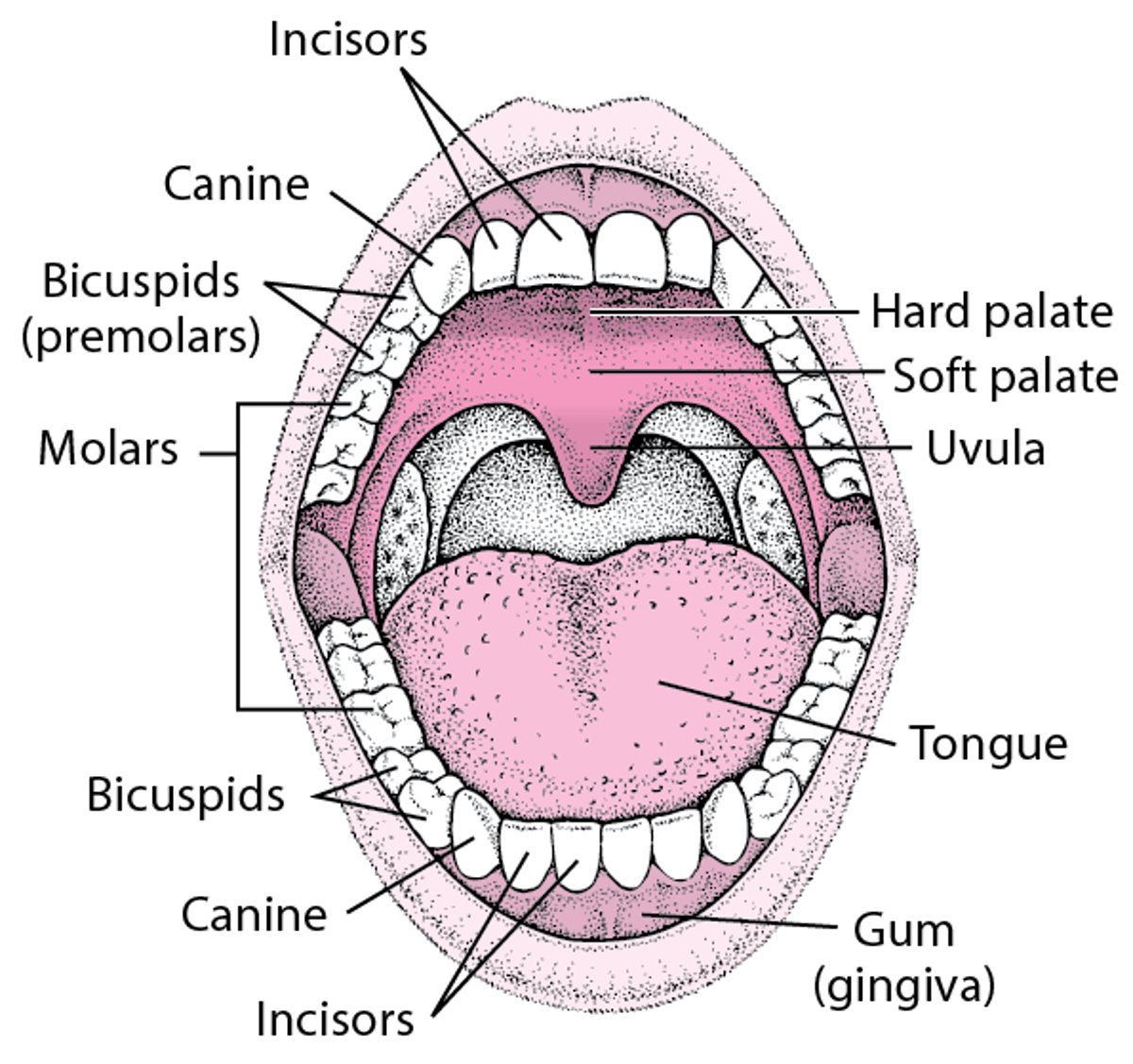
The palate, which is the roof of the mouth, is divided into two parts. The front part has ridges and is hard (hard palate). The back part is relatively smooth and soft (soft palate).
The moist mucous membranes lining the mouth continue outside, forming the pink and shiny portion of the lips, which meets the skin of the face at the vermilion border. The lip mucosa, although moistened by saliva, is prone to drying.
The uvula is a narrow muscular structure that hangs at the back of the mouth and can be seen when a person says "Ahh." The uvula hangs from the back of the soft palate, which separates the back of the nose from the back of the mouth. Normally, the uvula hangs vertically.
The tongue lies on the floor of the mouth and is used to taste and mix food. The tongue is not normally smooth. It is covered with tiny projections (papillae) that contain taste buds, some of which sense the taste of food.
The sense of taste is relatively simple, distinguishing sweet, sour, salty, bitter, and savory (also called umami, the taste of the flavoring agent monosodium glutamate). These tastes can be detected all over the tongue, but certain areas are more sensitive for each taste. Sweet detectors are located at the tip of the tongue. Salt detectors are located at the front sides of the tongue. Sour detectors are located along the sides of the tongue. Bitter detectors are located on the back one third of the tongue.
Smell is sensed by olfactory receptors high in the nose. The sense of smell is much more complex than that of taste, distinguishing many subtle variations. The senses of taste and smell work together to enable people to recognize and appreciate flavors (see Overview of Smell and Taste Disorders).
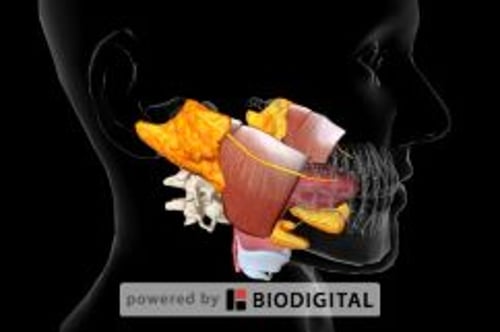
A View of the Mouth

The salivary glands produce saliva. There are three major pairs of salivary glands: parotid, submandibular, and sublingual. Besides the major salivary glands, many tiny salivary glands are distributed throughout the mouth. Saliva passes from the glands into the mouth through small tubes (ducts).
Saliva serves several purposes. Saliva aids in chewing and eating by gathering food into lumps so that food can slide out of the mouth and down the esophagus and by dissolving foods so that they can more easily be tasted. Saliva also coats food particles with digestive enzymes and begins digestion. After food is eaten, the flow of saliva washes away bacteria that can cause tooth decay (cavities) and other disorders. Saliva helps keep the lining of the mouth healthy and prevents loss of minerals from teeth. It not only neutralizes acids produced by bacteria but also contains many substances (such as antibodies and enzymes) that kill bacteria, yeasts, and viruses.