


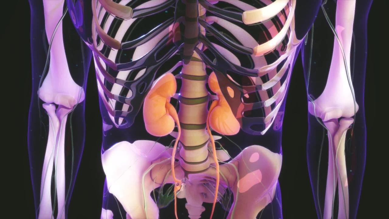
Renal (kidney) cortical necrosis is death of the tissue in the outer part of the kidney (cortex) that results from blockage of the small arteries that supply blood to the cortex and that causes acute kidney injury.
Usually the cause is a major, catastrophic disorder that decreases blood pressure.
Symptoms may include dark urine, decreased urine volume, fever, and pain in the side of the body.
Sometimes an imaging test or tissue analysis (biopsy) is done to confirm the diagnosis.
(See also Overview of Blood Vessel Disorders of the Kidneys.)
Renal cortical necrosis can occur at any age. About 10% of the cases occur in infants and children.
Causes of Cortical Necrosis of the Kidneys
In newborns, more than half of the cases occur after delivery complicated by premature detachment of the placenta. The next most common cause is a bacterial infection of the bloodstream (sepsis).
In children, renal cortical necrosis may occur after severe infection, severe dehydration, shock, or the hemolytic-uremic syndrome.
In women, about half of the cases occur after complications of pregnancy, such as premature detachment of the placenta or abnormal position of the placenta, bleeding from the uterus, infections immediately after childbirth, blockage of arteries by amniotic fluid, death of the fetus within the uterus, and preeclampsia.
Other causes in adults include severe infection, blood loss after injuries, rejection of a transplanted kidney, burns, inflammation of the pancreas (pancreatitis), snakebite, use of certain drugs, and poisoning caused by certain chemicals.
Symptoms of Cortical Necrosis of the Kidneys
The urine often becomes red or dark brown because of the presence of blood. Pain along both sides of the lower back may occur. A fever is often present. Changes in blood pressure, including mildly high pressure or even low pressure, are common. Urine flow may slow or stop.
Diagnosis of Cortical Necrosis of the Kidneys
Routine blood and urine tests
Imaging test
Sometimes kidney biopsy
Doctors may have difficulty making a diagnosis of renal cortical necrosis because it may resemble other types of acute kidney injury. Doctors may suspect renal cortical necrosis based on symptoms and the results of routine blood and urine tests in people who have predisposing conditions. The diagnosis is often confirmed with an imaging test such as ultrasonography or computed tomography angiography (CT angiography). Kidney biopsy can give doctors the most accurate diagnostic information, but a biopsy involves removing tissue and can cause complications and may be unnecessary if the diagnosis is evident. Thus, a biopsy is not done in most people.
Treatment of Cortical Necrosis of the Kidneys
Supportive care
Treatment of the underlying disorder
Treatment is supportive care, which may involve giving intravenous fluids, blood transfusion, antibiotics, dialysis, or a combination. The disorder that caused cortical necrosis is treated when possible.
In recent years, with improved treatment, prognosis has improved. About 80% of people live a year or longer, although most people need permanent dialysis or kidney transplantation.
More Information
The following English-language resources may be useful. Please note that THE MANUAL is not responsible for the content of these resources.
American Association of Kidney Patients (AAKP): AAKP improves the lives of patients through education, advocacy, and promotion of a sense of community among patients with kidney disease.
American Kidney Fund (AKF): AKF provides information about kidney disease and kidney transplant, needs-based financial assistance to help manage medical expenses, webinars for medical professionals, and opportunities for advocacy.
National Kidney Foundation (NKF): This clearinghouse provides everything from information on the basics of kidney function to access to treatment and support for people with kidney disease, continuing medical education courses, and research opportunities and grant support for medical professionals.
National Institute of Diabetes and Digestive and Kidney Diseases (NIDDK): General information on kidney diseases, including research discoveries, statistics, and community health and outreach programs.


