


VIEW PROFESSIONAL VERSION
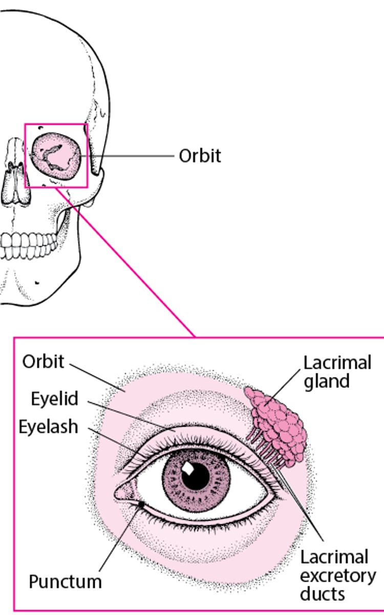
The eye sockets (orbits) are bony cavities that contain and protect the eyes and their supporting structures (see figures An Inside Look at the Eye and Structures That Protect the Eye). Disorders affecting the contents of the orbits include
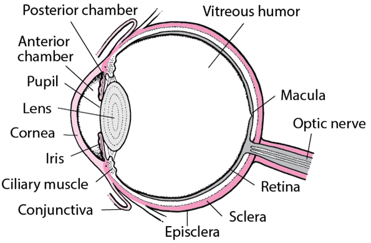
An Inside Look at the Eye

Infections (for example, orbital cellulitis and preseptal cellulitis)
Thyroid disease can also affect the contents of the orbits.
Many orbital disorders require treatment by a medical doctor who specializes in eye disorders (ophthalmologist).
Structures That Protect the Eye


Test your KnowledgeTake a Quiz!


