


Systemic lupus erythematosus is a chronic, multisystem, inflammatory disorder of autoimmune etiology, occurring predominantly in young women. Common manifestations may include arthralgias and arthritis, Raynaud syndrome, malar and other rashes, pleuritis or pericarditis, renal or central nervous system involvement, and autoimmune cytopenias. Diagnosis requires clinical and serologic criteria. Treatment of severe, ongoing, active disease requires corticosteroids and immunosuppressants.
Of all cases, 70 to 90% occur in women (usually of child-bearing age). Systemic lupus erythematosus (SLE) is more common and severe among Black and Asian patients than among White patients. It can affect patients of any age, including neonates. In some countries, the prevalence of SLE rivals that of rheumatoid arthritis
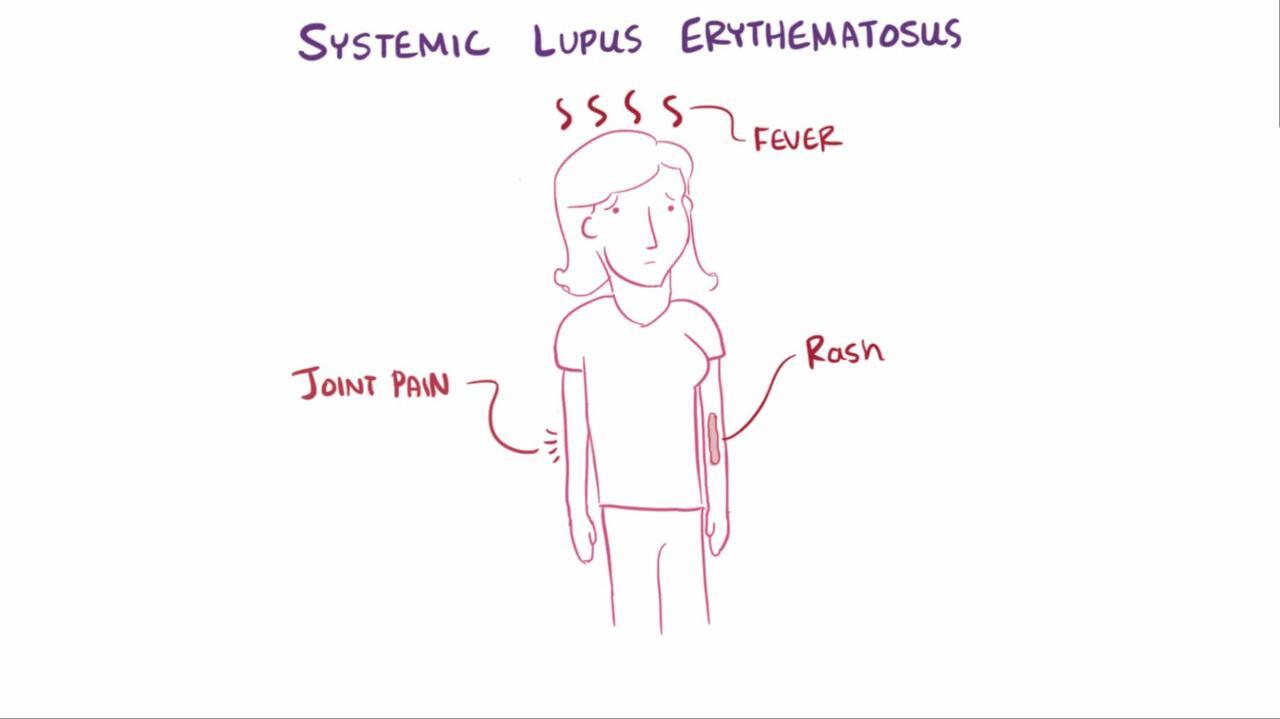
Symptoms and Signs of SLE
Clinical findings vary greatly. SLE may develop abruptly with fever or insidiously over months or years with episodes of arthralgias and malaise. Vascular headaches, epilepsy, or psychoses may be initial findings. Manifestations referable to any organ system may appear. Periodic exacerbations (flares) may occur.
Joint manifestations
Joint symptoms, ranging from intermittent arthralgias to acute polyarthritis, occur in about 90% of patients and may precede other manifestations by years. Most lupus polyarthritis is nondestructive and nondeforming. However, in long-standing disease, deformities without bone erosions may develop (eg, the metacarpophalangeal and interphalangeal joints may rarely develop reducible ulnar drift or swan-neck deformities without bony or cartilaginous erosions [Jaccoud arthritis]). As in many other chronic diseases, the prevalence of fibromyalgia is increased, which may cause diagnostic confusion in patients with periarticular and generalized pain and fatigue.
Skin and mucous membrane manifestations
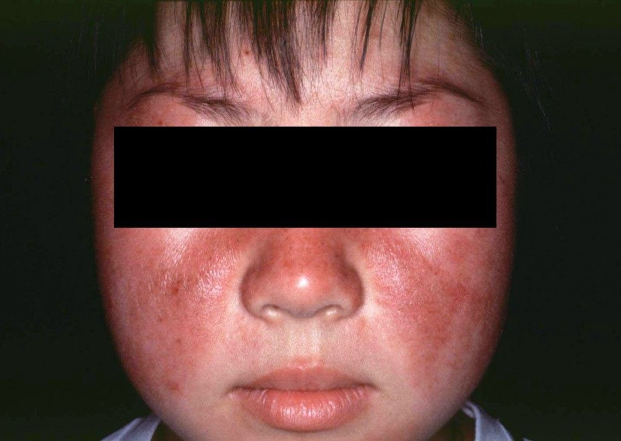
Skin lesions include malar butterfly erythema (flat or raised) that generally spares the nasolabial folds. The absence of papules and pustules and presence of skin atrophy help distinguish SLE from rosacea. A variety of other erythematous, firm, maculopapular lesions can occur elsewhere, including exposed areas of the face and neck, upper chest, and elbows. Skin blistering and ulceration are rare, although recurrent ulcers on mucous membranes (particularly the central portion of the hard palate near the junction of the hard and soft palate, the buccal and gum mucosa, and the anterior nasal septum) are common (sometimes called mucosal lupus); findings can sometimes mimic toxic epidermal necrolysis.

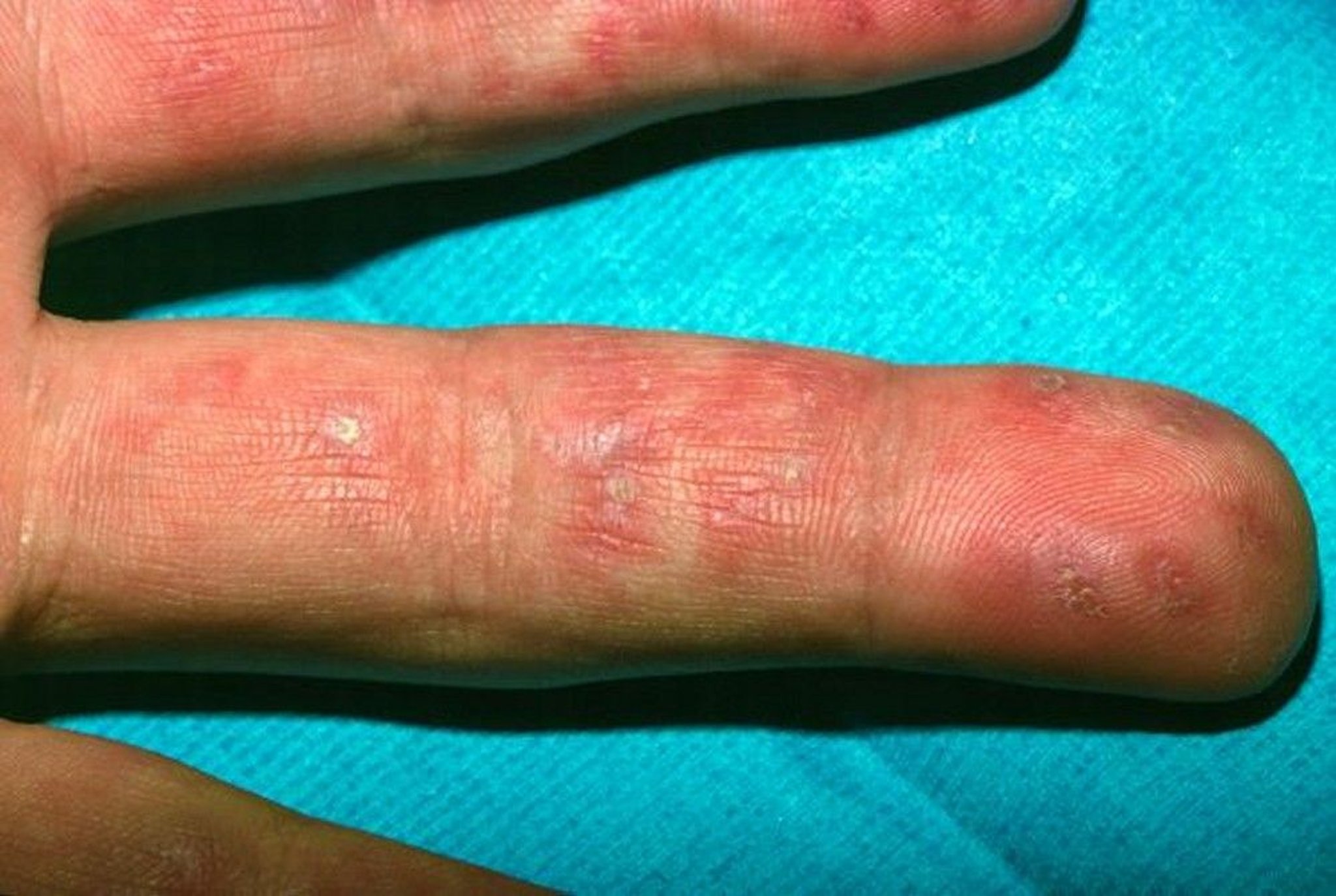
Image courtesy of Karen McKoy, MD.
Generalized or focal alopecia is common during active phases of SLE. Panniculitis can cause subcutaneous nodular lesions (sometimes called lupus panniculitis or profundus). Vasculitic skin lesions may include mottled erythema on the palms and fingers, periungual erythema, nail-fold infarcts, urticaria, and palpable purpura. Petechiae may develop secondary to thrombocytopenia. Photosensitivity occurs in some patients.
Lupus erythematosus tumidus is characterized by pink to violaceous urticarial nonscarring plaques and/or nodules, some annular, in light-exposed areas.

© Springer Science+Business Media
Chilblain lupus is characterized by tender, bright red to reddish blue nodules on the toes, fingers, nose, or ears that occur in cold weather. Some patients with SLE have features of lichen planus.
Raynaud syndrome due to vasospasm in the fingers and toes causes characteristic blanching and cyanosis.
(See also Variant Forms of Lupus.)
Cardiopulmonary manifestations
Cardiopulmonary symptoms commonly include recurrent pleurisy, with or without pleural effusion. Pneumonitis is rare, although minor impairments in pulmonary function are common. Diffuse alveolar hemorrhage occasionally occurs. Prognosis has traditionally been poor. Other complications include pulmonary emboli, pulmonary hypertension, and shrinking lung syndrome. Cardiac complications include pericarditis (most commonly) and myocarditis. Serious, rare complications are coronary artery vasculitis, valvular involvement, and Libman-Sacks endocarditis. Accelerated atherosclerosis is an increasing cause of morbidity and mortality. Congenital heart block can develop in neonates whose mother has the antibodies against Ro (SSA) or La (SSB).
Lymphoid tissue
Generalized adenopathy is common, particularly among children, young adults, and Black people; however, mediastinal adenopathy is not common. Splenomegaly occurs in 10% of patients.
Neurologic manifestations
Neurologic symptoms can result from involvement of any part of the central or peripheral nervous system or meninges. Mild cognitive impairment is common. There may also be headaches, personality changes, ischemic stroke, subarachnoid hemorrhage, seizures, psychoses, aseptic meningitis, peripheral and cranial neuropathies, transverse myelitis, choreoathetosis, or cerebellar dysfunction.
Renal manifestations
Renal involvement can develop at any time and may be the only manifestation of SLE (see Lupus Nephritis). It may be benign and asymptomatic or progressive and fatal. Renal lesions can range in severity from a focal, usually benign, glomerulitis to a diffuse, potentially fatal, membranoproliferative glomerulonephritis. Common manifestations include proteinuria (most often), an abnormal urinary sediment manifested by red blood cell casts and leukocytes, hypertension, and edema. Early lupus glomerulonephritis may be misdiagnosed as asymptomatic urinary tract infection.
Obstetric manifestations
Obstetric manifestations include early and late fetal loss. In patients with antiphospholipid antibodies, the risk of recurrent miscarriages is increased. Pregnancy can be successful (see SLE in Pregnancy), particularly after 6 to 12 months of remission, but SLE flares are common during pregnancy and especially during the postpartum period. Pregnancy should be timed for when disease is in remission. During pregnancy, the patient should be monitored closely for any disease flare or thrombotic events by a multidisciplinary team that includes an obstetrician who specializes in high-risk pregnancies. Women who are SSA antibody-positive should have weekly fetal ultrasonography between week 18 and week 26 to assess for congenital heart block.
Hematologic manifestations
Hematologic manifestations include anemia (anemia of chronic disease, autoimmune hemolytic anemia), leukopenia (usually lymphocytopenia, with < 1500 cells/mcL), and thrombocytopenia (usually mild but sometimes life-threatening autoimmune thrombocytopenia). Recurrent arterial or venous thrombosis, thrombocytopenia, and a high probability of obstetric complications occur in patients with antiphospholipid antibodies. Thromboses probably account for many of the complications of SLE, including obstetric complications. Macrophage activation syndrome can occur.
Gastrointestinal manifestations
Gastrointestinal manifestations can result from bowel vasculitis or impaired bowel motility. In addition, pancreatitis can rarely result from SLE. Manifestations may include abdominal pain resulting from serositis, nausea, vomiting, manifestations of bowel perforation, and pseudo-obstruction. SLE rarely causes parenchymal liver disease.
Diagnosis of SLE
Clinical criteria
Cytopenias
Autoantibodies
SLE should be suspected in patients, particularly young women, with any of the symptoms and signs. However, early-stage SLE can mimic other connective (or nonconnective) tissue disorders, including rheumatoid arthritis if arthritic symptoms predominate. Mixed connective tissue disease can mimic SLE but also may involve features of systemic sclerosis, rheumatoid-like polyarthritis, and polymyositis. Infections (eg, bacterial endocarditis, histoplasmosis) can mimic SLE and may develop as a result of treatment-caused immunosuppression. Disorders such as sarcoidosis and paraneoplastic syndromes can also mimic SLE.
Laboratory testing may differentiate SLE from other connective tissue disorders. Routine testing should include the following:
Antinuclear antibodies (ANA) and anti–double-stranded (ds) DNA (anti-dsDNA)
Complete blood count (CBC)
Urinalysis
Chemistry profile including renal and liver enzymes
In clinical practice, some clinicians rely on the classification criteria for SLE developed by the European League Against Rheumatism/American College of Rheumatology (EULAR/ACR; see table EULAR/ACR Criteria for the Classification of Systemic Lupus Erythematosus). Patients are eligible for these criteria only if they have a positive ANA ≥ 1:80. The 2019 EULAR/ACR classification criteria include clinical and immunologic domains, and each criterion is assigned a weight of 2 to 10. If the patient's score is 10 or more, and at least one clinical criterion is fulfilled, disease is classified as SLE. However, a positive ANA does not indicate a diagnosis of lupus. A positive ANA test in the presence of fatigue and generalized myofascial pain without other clinical or laboratory findings is rarely significant.
Fluorescent ANA
The fluorescent test for ANA is the best initial test for SLE in patients who have compatible symptoms and signs; positive ANA tests (usually in high titer: > 1:80) occur in >
Other ANA and anticytoplasmic antibodies
The ANA test is very sensitive, but it is not specific for SLE; thus, evidence of other autoantibodies is used to aid in diagnosis. They include Ro (SSA), La (SSB), Smith (Sm), ribonucleoprotein (RNP), and dsDNA. Ro is predominantly cytoplasmic; anti-Ro antibodies are occasionally present in ANA-negative SLE patients presenting with chronic cutaneous lupus. Anti-Ro is the causal antibody for neonatal lupus and congenital heart block. Anti-Sm is highly specific for SLE but, like anti-dsDNA, is not sensitive. Anti-RNP occurs in patients with SLE, mixed connective tissue disease, and occasionally other systemic autoimmune disorders and systemic sclerosis.
Other blood tests
Leukopenia (usually lymphopenia) is common. Hemolytic anemia may occur, but low hemoglobin and red blood cell counts are more often due to the anemia of chronic disease. Thrombocytopenia in SLE may be difficult or impossible to differentiate from idiopathic thrombocytopenic purpura except that patients have other features of SLE and/or SLE-specific antibodies (anti-dsDNA or anti-Sm). False-positive serologic tests for syphilis occur in 5 to 10% of SLE patients. These test results may be associated with the lupus anticoagulant and a prolonged partial thromboplastin time (PTT). Abnormal values in one or more of these assays suggest the presence of antiphospholipid antibodies (eg, anticardiolipin antibodies), which should then be measured directly by enzyme-linked immunosorbent assay (ELISA). Antiphospholipid antibodies are associated with arterial or venous thrombosis, thrombocytopenia, and, during pregnancy, spontaneous abortion or late fetal death but may be present in asymptomatic patients.
Other tests help monitor disease severity and determine the need for treatment. Serum complement levels (C3, C4) are often depressed in active disease and are usually lowest in patients with active nephritis. Erythrocyte sedimentation rate (ESR) is elevated frequently during active disease. C-reactive protein levels are not necessarily elevated; high levels raise the concern for infection and/or serositis.
Renal involvement
Screening for renal involvement begins with urinalysis. Red blood cell (RBC) and/or white blood cell casts suggest active nephritis. Urinalysis should be done at regular intervals (eg, every 3 to 6 months), even for patients in apparent remission, because kidney disease is usually asymptomatic. Proteinuria can be estimated by the urine protein/creatinine ratio or measured in a 24-hour urine collection. Renal biopsy is indicated in patients whose protein excretion is > 500 mg/day and who have hematuria (thought to be glomerular) or RBC casts and is helpful in evaluating the status of renal disease (ie, active inflammation vs postinflammatory scarring) and in guiding therapy. Patients with chronic renal insufficiency and mostly sclerotic glomeruli are not likely to benefit from aggressive immunosuppressive therapy.
Prognosis for SLE
The course is usually chronic, relapsing, and unpredictable. Remissions may last for years. If the initial acute phase is controlled, even if very severe (eg, with cerebral thrombosis or severe nephritis), the long-term prognosis is usually good. The 10-year survival in most developed countries is > 95%. Improved prognosis is in part due to earlier diagnosis and more effective therapies. Complications include infection from immunosuppression or osteoporosis from long-term corticosteroid use. Increased risk of coronary artery disease can contribute to premature death.
Treatment of SLE
Nonsteroidal anti-inflammatory drugs (NSAIDs) in addition to antimalarials for mild disease
Corticosteroids, immunosuppressants, and antimalarials for severe disease
To simplify therapy, SLE should be classified as mild to moderate (eg, fever, arthritis, pleurisy, pericarditis, rash) or severe (eg, hemolytic anemia, severe thrombocytopenic purpura, massive pleural and pericardial involvement, diffuse alveolar hemorrhage or pneumonitis, nephritis, acute vasculitis of the extremities or gastrointestinal tract, florid central nervous system [CNS] involvement).
1glucose-6-phosphate dehydrogenase (G6PD) deficiency because it can cause hemolysis.
(See also recommendations for the management of SLE from the European League Against Rheumatism [EULAR].)
Mild to moderate disease
Severe disease
Treatment includes induction therapy to control acute severe manifestations followed by maintenance therapy. Corticosteroids are first-line therapy. A combination of a corticosteroid and other immunosuppressants is typically used in active severe disease (ie, lupus nephritis with impaired renal function or CNS involvement).
The complication for which there is the strongest evidence for treatment efficacy is lupus nephritis
345). Classification of lupus nephritis is based on histologic findings on renal biopsy (see table Classification of Lupus Nephritis).
In CNS lupus, including transverse myelitis
2).
Patients with end-stage renal disease can undergo kidney transplantation, as an alternative to dialysis, with a successful outcome, especially if their disease has been in remission.
6).
Maintenance therapy
2). Treatment should be guided by clinical features primarily, although anti-dsDNA antibody titers or serum complement levels may be followed, particularly if they have correlated with disease activity in the past. However, anti-dsDNA antibody titers or serum complement levels may not parallel nonrenal disease flares. Other pertinent blood and urine tests may be used to assess specific organ involvement.
prevention of osteoporosis) should be considered in patients taking corticosteroids long term.
If combination immunosuppressive therapy is used, patients should be given prophylaxis for opportunistic infections, such as Pneumocystis jirovecii (see prevention of Pneumocystis jirovecii pneumonia), and vaccines against common infections (eg, streptococcal pneumonia, influenza, COVID-19).
Coexisting medical conditions and pregnancy
All patients should be closely monitored for atherosclerosis, and cardiovascular risk reduction is a key part of management (see treatment of atherosclerosis). Long-term anticoagulation is vital in patients with antiphospholipid antibodies and history of thrombosis (see also Anticoagulants).
Treatment references
1. Alarcón GS, McGwin G, Bertoli AM, et alAnn Rheum Dis 66(9):1168–1172, 2007. doi: 10.1136/ard.2006.068676
2. Fanouriakis A, Kostopoulou M, Alunno A, et al: 2019 update of the EULAR recommendations for the management of systemic lupus erythematosus. Ann Rheum Dis 78(6):736-745, 2019. doi:10.1136/annrheumdis-2019-215089
3. Furie R, Rovin BH, Houssiau F, et alN Engl J Med 383(12):1117-1128, 2020. doi:10.1056/NEJMoa2001180
4. Rovin BH, Teng YKO, Ginzler EM, et al: Efficacy and safety of voclosporin versus placebo for lupus nephritis (AURORA 1): a double-blind, randomised, multicentre, placebo-controlled, phase 3 trial.Lancet 397(10289):2070-2080, 2021. doi:10.1016/S0140-6736(21)00578-X. Erratum in: Lancet 397(10289):2048, 2021.
5. Bajema IM, Wilhelmus S, Alpers CE, et al. Revision of the International Society of Nephrology/Renal Pathology Society classification for lupus nephritis: clarification of definitions, and modified National Institutes of Health activity and chronicity indices. Kidney Int 93(4):789-796, 2018. doi:10.1016/j.kint.2017.11.023
6. Morand EF, Furie R, Tanaka Y, et alN Engl J Med 382(3):211-221, 2020. doi:10.1056/NEJMoa1912196
Key Points
Joint and skin manifestations are classic in SLE, but the disorder can affect various organ systems, such as the skin, heart and lungs, lymphoid tissue, kidneys, and gastrointestinal, hematologic, reproductive, and nervous systems.
The European League Against Rheumatism/American College of Rheumatology (EULAR/ACR) criteria can be used to confirm the diagnosis of SLE.
Among tests, use the highly sensitive ANA for screening, but use more specific autoantibodies (eg, anti-dsDNA, anti-Sm) for confirmation.
Evaluate all patients for kidney involvement.
Use corticosteroids at the lowest possible dose and other drugs that control inflammation to maintain remission.
More Information
The following English-language resources may be useful. Please note that THE MANUAL is not responsible for the content of these resources.


