Select an Online Manual
THE MERCK MANUAL MEDICAL LIBRARY: The Merck Manual of Diagnosis and Therapy
Tips for better results
ABCDEFGHI
JKLMNOPQR
STUVWXYZ
In This Topic
Introduction
Back to Top

Section

Subject

Topics

Introduction

Update Me

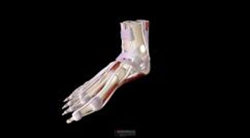
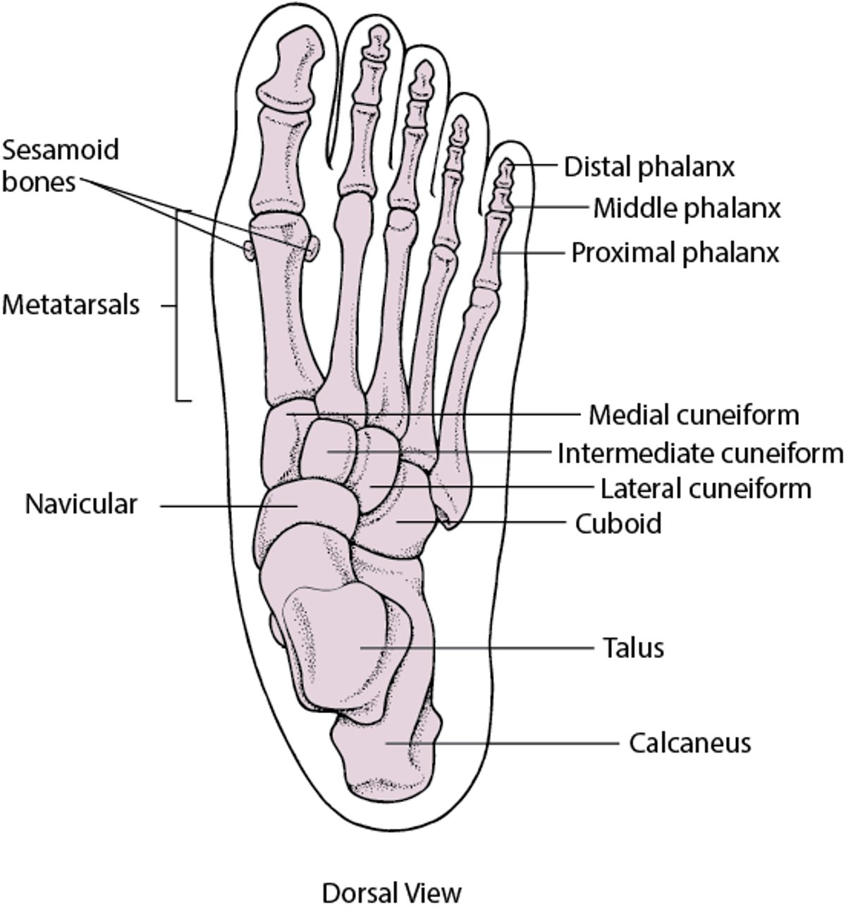
Most foot problems result from anatomic disorders or abnormal function of articular or extra-articular structures (see Fig. 1: Foot and Ankle Disorders: Bones of the foot.Figures). Less commonly, foot problems reflect a systemic disorder (see Table 1: Foot and Ankle Disorders: Foot Manifestations of Systemic DisordersTables).

Fig. 1

Bones of the foot.

Bones of the foot.

Table 1

Foot Manifestations of Systemic Disorders

Foot Symptoms or Signs

Possible Cause

Pain at rest (feet elevated), relieved by dependency

End-stage peripheral arterial disease

Cold, red, or cyanotic feet

Advanced arterial ischemia

Episodically red, hot, very painful feet

Erythromelalgia—idiopathic (most commonly) or secondary to various disorders (eg, myeloproliferative disorders, which are rare)

Foot pain that becomes severe within seconds or possibly minutes, particularly in patients with atrial fibrillation; foot often cool

Embolic arterial occlusion

Cyanosis of a single toe (blue toe syndrome)

Thromboembolic disease due to aortic-iliac stenosis, arrhythmia, or cholesterol embolization (after coronary artery bypass or catheterization); warfarin Some Trade Names
COUMADIN
Click for Drug Monograph
therapy

Bilateral episodic digital discomfort, pallor, and cyanosis

Raynaud's disease or phenomenon

Bilateral painless cyanosis (in a young female)

Acrocyanosis

Bilateral edema

Renal, hepatic, or cardiac dysfunction; drugs (eg, Ca channel blockers)

Unilateral edema

Deep vein thrombosis, lymphatic obstruction

Firm non-pitting foot and leg edema

Lymphedema, scleroderma

Firm non-pitting edema with nodular appearance above the malleoli

Pretibial myxedema

Edema with hemosiderin deposition and brownish discoloration

Venous insufficiency, recurrent small-vessel vasculitis

Edema of feet and toes, numbness and pain at the ankle and heel (tarsal tunnel syndrome)

Hypothyroidism, relapsing symmetric seronegative synovitis (rare)

Red, dusky patches on the dorsum with flaccid bullae (necrolytic acral erythema)

Hepatitis C, vasculitis, emboli

Isolated toe swelling and deformity with pain (sausage digits)

Psoriatic arthritis, reactive arthritis, other spondyloarthropathies; crystal-induced arthritis,

Painful feet with paresthesias

Peripheral neuropathy (local or systemic—eg, diabetic neuropathy)

Pain or paresthesias in the leg and foot; pain in the foot and back when the leg is extended, relieved when the knee is flexed

Sciatica

Toe, foot, or ankle pain with warmth and redness

Gout, stress fracture

Thickened (> 22 mm) heel pad as determined with radiographic or ultrasonic imaging techniques

Hyperpituitarism with acromegaly

In people with diabetes and people with peripheral vascular disease, careful examination of the feet, with evaluation of vascular sufficiency and neurologic integrity, should be done at least twice/year. People with these diseases should examine their own feet at least once/day.

The feet are also common sites for corns and calluses (see Cornification Disorders) and infections by fungus (see Fungal Skin Infections: Tinea Pedis), bacteria (see Bacterial Skin Infections), and viruses (see Viral Skin Diseases).

Table 2: Foot and Ankle Disorders: Common Foot and Ankle Disorders by Anatomic SiteTables lists foot and ankle disorders according to anatomic site. Table 3: Foot and Ankle Disorders: Disorders Associated With Heel Pain According to LocationTables lists common causes of heel pain according to location.

Table 2

Common Foot and Ankle Disorders by Anatomic Site

Ankle (anterolateral)

Meniscoid body

Neuralgia of the intermediate dorsal cutaneous nerve

Peroneal tenosynovitis

Ankle (medial)

Tarsal tunnel syndrome

Tibialis posterior tendinosis

Ball of the foot

Corns and calluses

Freiberg's disease

Interdigital nerve pain (Morton's neuroma)

Metatarsophalangeal joint pain

Sesamoiditis

Heel (plantar)

Inferior calcaneal bursitis

Plantar fasciosis

Heel (posterior)

Achilles tendon enthesopathy

Anterior Achilles tendon bursitis

Posterior Achilles tendon bursitis

Heel (sides)

Epiphysitis of the calcaneus (Sever's disease)

Medial plantar nerve entrapment

Plantar arch (sole)

Plantar fascial sprain

Plantar fibromatosis

Toe

Bunion

Hammer toe

Ingrown toenail

Onychomycosis

Paronychia

Table 3

Disorders Associated With Heel Pain According to Location

Location of Pain

Associated Disorder

Plantar surface of the heel

Plantar fasciosis (plantar fasciitis, calcaneal spur syndrome), inferior calcaneal bursitis

Medial and lateral margins of the heel

Epiphysitis of the calcaneus (Sever's disease, in children), medial plantar nerve entrapment, sometimes tarsal tunnel syndrome

Anterior to the Achilles tendon at the retromalleolar space

Fracture of the posterolateral talar tubercle, tibialis posterior tendinosis, anterior Achilles tendon bursitis, tarsal tunnel syndrome

Posterior to the Achilles tendon

Posterior Achilles tendon bursitis, tendon nodules

Calcaneal insertion of the Achilles tendon

Achilles tendon enthesopathy

Last full review/revision March 2008 by Kendrick Alan Whitney, DPM

Content last modified March 2008

Back to Top

Next: Achilles Tendon Enthesopathy

Audio
Figures
Photographs
Tables
Videos