


DR P. MARAZZI/SCIENCE PHOTO LIBRARY
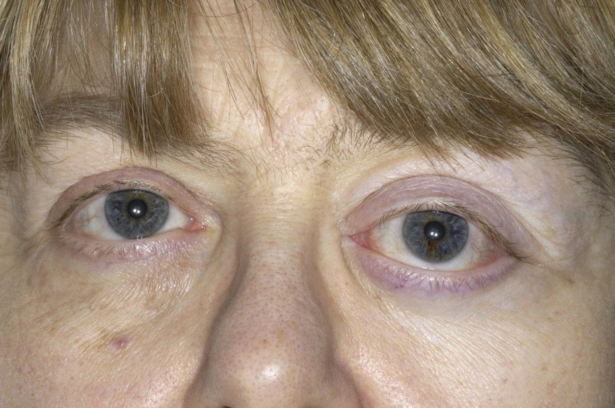
Proptosis is protrusion of the eyeball. Exophthalmos means the same thing, and this term is usually used when describing proptosis due to Graves disease. Disorders that may cause changes in the appearance of the face and eyes that resemble proptosis but are not include hyperthyroidism without infiltrative eye disease, Cushing disease, and severe obesity.
Etiology of Proptosis
The most common cause in adults is Graves disease (see table Some Causes of Proptosis), which causes edema and lymphoid infiltration of the orbital tissues.
The most common cause in children is orbital cellulitis.
Evaluation of Proptosis
Rate of onset may provide a clue to diagnosis. Sudden unilateral onset suggests intraorbital hemorrhage (which can occur after surgery, retrobulbar injection, or trauma) or inflammation of the orbit or paranasal sinuses. A 2- to 3-week onset suggests chronic inflammation or orbital inflammatory pseudotumor (non-neoplastic cellular infiltration and proliferation); slower onset suggests an orbital tumor.
Ocular examination findings typical of hyperthyroidism but unrelated to infiltrative eye disease include eyelid retraction, eyelid lag, temporal flare of the upper eyelid, and staring. Other signs include eyelid erythema and conjunctival hyperemia. Prolonged exposure of larger-than-usual areas of the eyeball to air causes corneal drying and can lead to infection and ulceration.
Red flags
The following findings are of particular concern:
Eye pain or redness
Headache
Loss of vision
Diplopia
Fever
Pulsating proptosis
Neonatal proptosis
Testing
Proptosis can be confirmed with exophthalmometry, which measures the distance between the lateral angle of the bony orbit and the cornea; normal values are often < 20 mm, though there is some variation by race, ethnicity, and gender. CT or MRI of the orbits is often useful to confirm the diagnosis and to identify structural causes of unilateral proptosis. Thyroid function testing is indicated when Graves disease is suspected.
Treatment of Proptosis
Key Points
The most common cause of bilateral proptosis in adults is Graves disease.
Acute unilateral proptosis suggests infection or vascular disorder (eg, hemorrhage, fistula, cavernous sinus thrombosis).
Chronic unilateral proptosis suggests tumor.
Do CT or MRI and thyroid function testing when Graves disease is suspected.
Apply lubrication to exposed cornea.


