


Seborrheic dermatitis is a common inflammatory condition of skin regions with a high density of sebaceous glands (eg, face, scalp, sternum). The cause is unknown, but species of Malassezia, a normal skin yeast, play an important role. This dermatitis occurs with increased frequency in patients with HIV and in those with certain neurologic disorders. Seborrheic dermatitis causes occasional pruritus, dandruff, and yellow, greasy scaling on the scalp, along the hairline, and on the face. Diagnosis is made by examination. Treatment is with antifungals, topical corticosteroids, tar, and keratolytics.
(See also Definition of Dermatitis.)
Despite the name, the composition and flow of sebum are usually normal. The pathogenesis of seborrheic dermatitis is unclear, but its activity has been linked to the number of Malassezia yeasts present on the skin and to the inflammatory reaction to them.
Seborrheic dermatitis occurs most often in infants, usually within the first 3 months of life, and in adults aged 30 to 70 years. The incidence and severity of disease seem to be affected by genetic factors, emotional or physical stress, and climate (usually worse in cold weather). Seborrheic dermatitis may precede or be associated with psoriasis (called seborrhiasis or sebopsoriasis). Seborrheic dermatitis may be more common and more severe among patients with neurologic disorders (especially Parkinson disease), because of, for example, changes in the activity of sebaceous glands, or among those with HIV/AIDS, likely because of an imbalance of T-cell pro- and anti-inflammatory responses. Very rarely, the dermatitis becomes generalized.
Symptoms and Signs of Seborrheic Dermatitis
Symptoms of seborrheic dermatitis develop gradually, and the dermatitis is usually apparent only as dry flakes or greasy diffuse scaling of the scalp (dandruff) with variable pruritus.
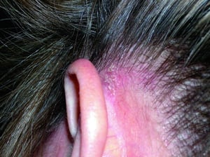
In severe disease, yellow-red scaling papules appear along the hairline, behind the ears, on the eyebrows, in the nasolabial folds, and over the sternum. Marginal blepharitis with dry yellow crusts and conjunctival irritation may develop. Seborrheic dermatitis does not cause hair loss.
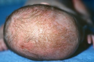
Newborns may develop seborrheic dermatitis with a thick, yellow, crusted scalp lesion (cradle cap); fissuring and yellow scaling behind the ears; red facial papules; and stubborn diaper rash.
Older children and adults may develop thick, tenacious, scaly plaques on the scalp that may measure 1 to 2 cm in diameter.
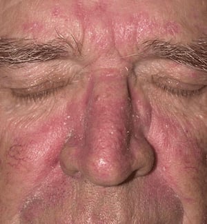
Image provided by Thomas Habif, MD.
© Springer Science+Business Media
Biophoto Associates/SCIENCE PHOTO LIBRARY


Image provided by Thomas Habif, MD.


© Springer Science+Business Media


Biophoto Associates/SCIENCE PHOTO LIBRARY
Diagnosis of Seborrheic Dermatitis
Clinical evaluation
Diagnosis of seborrheic dermatitis is made by physical examination.
Seborrheic dermatitis of the scalp must be differentiated from other disorders:
Atopic dermatitis of the scalp: This disorder typically first manifests with fine, white, dry scaling rather than the greasy yellowish scale of seborrheic dermatitis.
Scalp psoriasis: The erythematous and scaly plaques are sharply demarcated.
Rosacea: When rosacea affects the face, it first manifests with erythema, papules, and papulopustules but not with scaling (however, patients can have both seborrheic dermatitis and rosacea).
Treatment of Seborrheic Dermatitis
Topical therapy with antifungals, corticosteroids, tar, keratolytics, and calcineurin inhibitors
Adults and older children
Treatment of seborrheic dermatitis of the scalp should include shampooing at least twice a week, because less frequent shampooing enables proliferation of Malassezia
Infants and children
Key Points
In adults, seborrheic dermatitis causes dandruff and sometimes scaling on the scalp, around the eyebrows, nasolabial folds, nose, external auditory canal, behind the ears, and on the sternum.
Seborrheic dermatitis can cause a thick, yellow, crusted scalp lesion in newborns or thick, scaly scalp plaques in older children and adults.
Treatments include topical antifungals; antifungal, keratolytic, and tar shampoos; and topical corticosteroids.


