


Malignant melanoma arises from melanocytes in a pigmented area (eg, skin, mucous membranes, eyes, or central nervous system). Metastasis is correlated with depth of dermal invasion. With metastasis, prognosis is poor. Diagnosis is by biopsy. Wide surgical excision is the rule for operable tumors. Metastatic disease requires systemic therapy but is difficult to cure.
(See also Overview of Skin Cancer.)
In 2023, approximately 97,610 new cases of melanoma are estimated to occur in the United States, causing an estimated 7,990 deaths (1). Lifetime risk is approximately 2.6% for White people, 0.1% for Black people, and 0.6% for Hispanic people. Melanoma accounts for < 2% of total skin cancers diagnosed in the United States but causes most skin cancer deaths.
Melanomas occur mainly on the skin but also on the mucosa of the oral, genital, and rectal regions and conjunctiva. Melanomas may also develop in the choroid layer of the eye, in the leptomeninges (pia or arachnoid mater), and in the nail beds.


Melanomas vary in size, shape, and color (usually pigmented) and in their propensity to invade and metastasize. Metastasis occurs via lymphatics and blood vessels. Local metastasis results in the formation of nearby satellite papules or nodules that may or may not be pigmented. Metastasis to skin or internal organs may occur, and, occasionally, metastatic nodules or enlarged lymph nodes are discovered before the primary lesion is identified.

© Springer Science+Business Media
General reference
1. American Cancer Society: Key Statistics for Melanoma Skin Cancer. Atlanta, American Cancer Society, 2023.
Risk Factors for Melanoma
Risk factors for melanoma include
Sun exposure, particularly repeated blistering sunburns
Repeated tanning with ultraviolet A (UVA) or psoralen plus UVA (PUVA) treatments
Nonmelanoma skin cancer
Family and personal history of melanoma
Light skin, freckling
Atypical moles, particularly > 5
Increased numbers of melanocytic nevi
Immunosuppression
Lentigo maligna
Congenital melanocytic nevus > 20 cm (giant congenital nevi)
Atypical mole syndrome (dysplastic nevus syndrome)
Familial atypical mole–melanoma syndrome
Germline mutations in oncogenes, including BRCA2
Advanced age


Patients with a personal history of melanoma have an increased risk of additional melanomas. People who have one or more first-degree relatives with a history of melanoma have an increased risk (up to 6 or 8 times) over those without a family history.
Atypical mole syndrome is the presence of large numbers of moles (eg,> 50), at least one of which is atypical and at least one of which is > 8 mm in diameter.
Familial atypical mole–melanoma syndrome is the presence of multiple atypical moles and melanoma in 2 or more first-degree relatives; such people are at markedly increased risk (25 times) of melanoma.
Melanoma is less common among people with dark skin; when it occurs in people with dark skin, the nail beds, palms, and soles are more often affected.
Approximately 30% of melanomas develop from pigmented moles (about half each from typical and atypical moles); almost all the rest arise from melanocytes in normal skin. Atypical moles (dysplastic nevi) may be precursors to melanoma.
Although melanomas occur during pregnancy, pregnancy does not increase the likelihood that a mole will become a melanoma; moles frequently change in size and darken uniformly during pregnancy.
The very rare melanomas of childhood almost always arise in the leptomeninges or from giant congenital nevi.
In all people, lesions that have certain characteristics of concern, such as size, irregular borders, recent enlargement, darkening, ulceration, or bleeding, should be evaluated (see diagnosis of melanoma).

MID ESSEX HOSPITAL SERVICES NHS TRUST / SCIENCE PHOTO LIBRARY
Classification of Melanoma
There are 4 main types of melanoma and a few minor subtypes.
Superficial spreading melanoma
This type accounts for 70% of melanomas. Typically asymptomatic, it occurs most commonly on women’s legs and men’s torsos.
The lesion is usually a plaque with irregular, raised, indurated, and tan or brown areas, which often have red, white, black, and blue spots or small, sometimes protuberant blue-black nodules. Small notchlike indentations of the margins may be noted, along with enlargement or color change.
Histologically, atypical melanocytes characteristically invade the dermis and epidermis. This type of melanoma most commonly has activating mutations in the BRAF gene at V600.


Nodular melanoma
This type accounts for 15 to 30% of melanomas. It may occur anywhere on the body as a dark, protuberant papule or a plaque that varies from pearl to gray to black. Occasionally, a lesion contains little if any pigment or may look like a vascular tumor.
Unless it ulcerates, nodular melanoma is asymptomatic, but patients usually seek advice because the lesion enlarges rapidly.


Lentigo maligna melanoma
This type accounts for 5% of melanomas. It tends to occur in older adults. It arises from lentigo maligna (Hutchinson freckle or malignant melanoma in situ—a frecklelike tan or brown macule).
Lentigo maligna usually occurs on the face or other areas of chronic sun exposure as an asymptomatic, flat, tan or brown, irregularly shaped macule or patch with darker brown or black spots scattered irregularly on its surface. In lentigo maligna, both normal and malignant melanocytes are confined to the epidermis.
When malignant melanocytes invade the dermis, the lesion is called lentigo maligna melanoma, and the cancer may metastasize.
This type of melanoma most commonly has mutations in the C-kit gene.


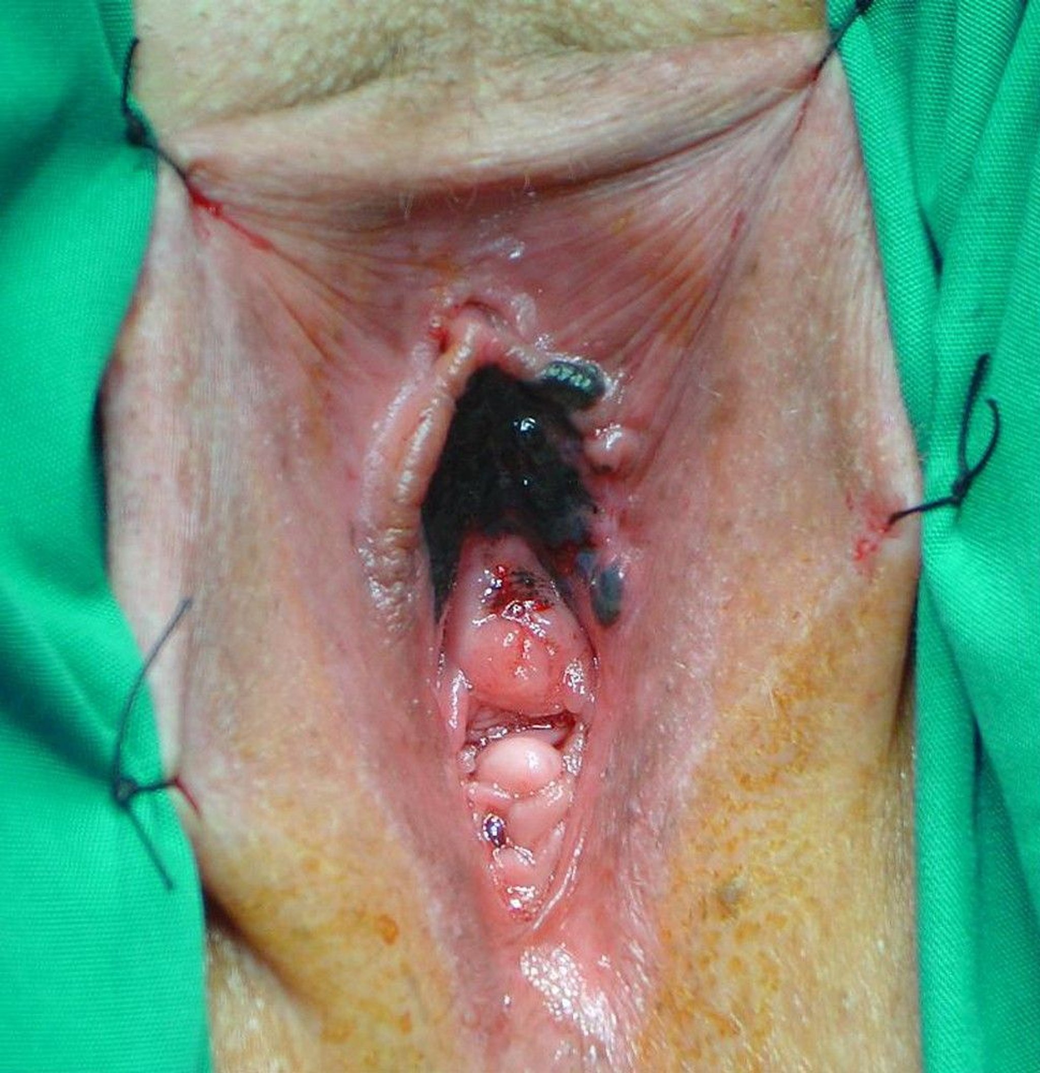
Acral-lentiginous melanoma
This type accounts for only 2 to 10% of melanomas. Incidence is probably the same regardless of skin pigmentation, but because people with dark skin infrequently develop other forms of melanoma, acral-lentiginous melanoma is the most common type among them.
Acral-lentiginous melanoma arises on palmar, plantar, and subungual skin and has a characteristic histologic picture similar to that of lentigo maligna melanoma.
This type of melanoma often has mutations in the C-kit gene.


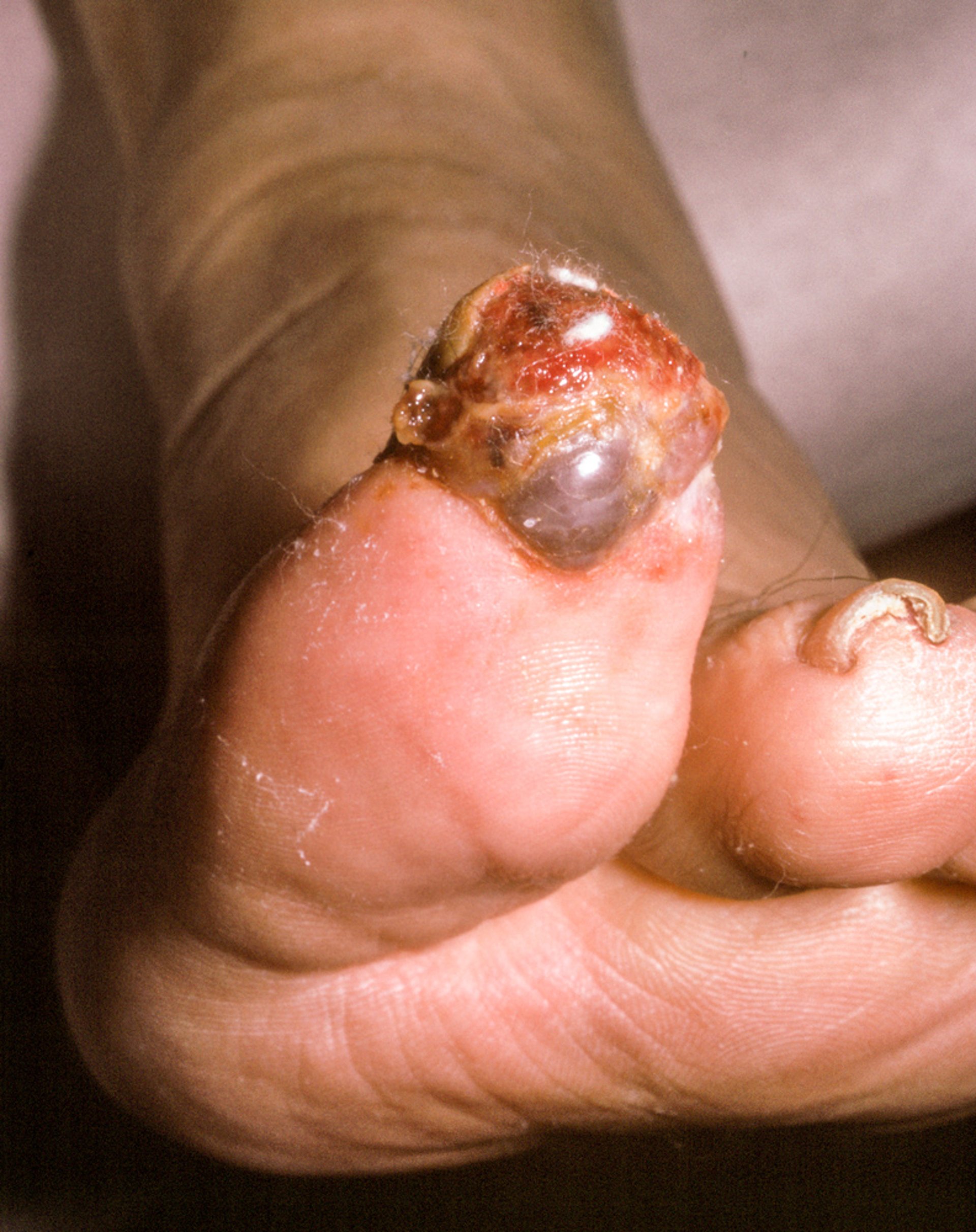
Photo courtesy of Karen McKoy, MD.
Amelanotic melanoma
Amelanotic melanoma is a type of melanoma that does not produce pigment. It can be any of the 4 main types and is most often grouped with the minor categories of melanoma such as spitzoid melanoma, desmoplastic melanoma, neurotropic melanoma, and others.
Accounting for < 10% of melanomas, amelanotic melanomas may be pink, red, or slightly light brown and may have well-defined borders. Their appearance may suggest benign lesions, or a form of nonmelanoma skin cancer, and thereby lead to a late diagnosis and possibly a worse prognosis.


Diagnosis of Melanoma
Biopsy
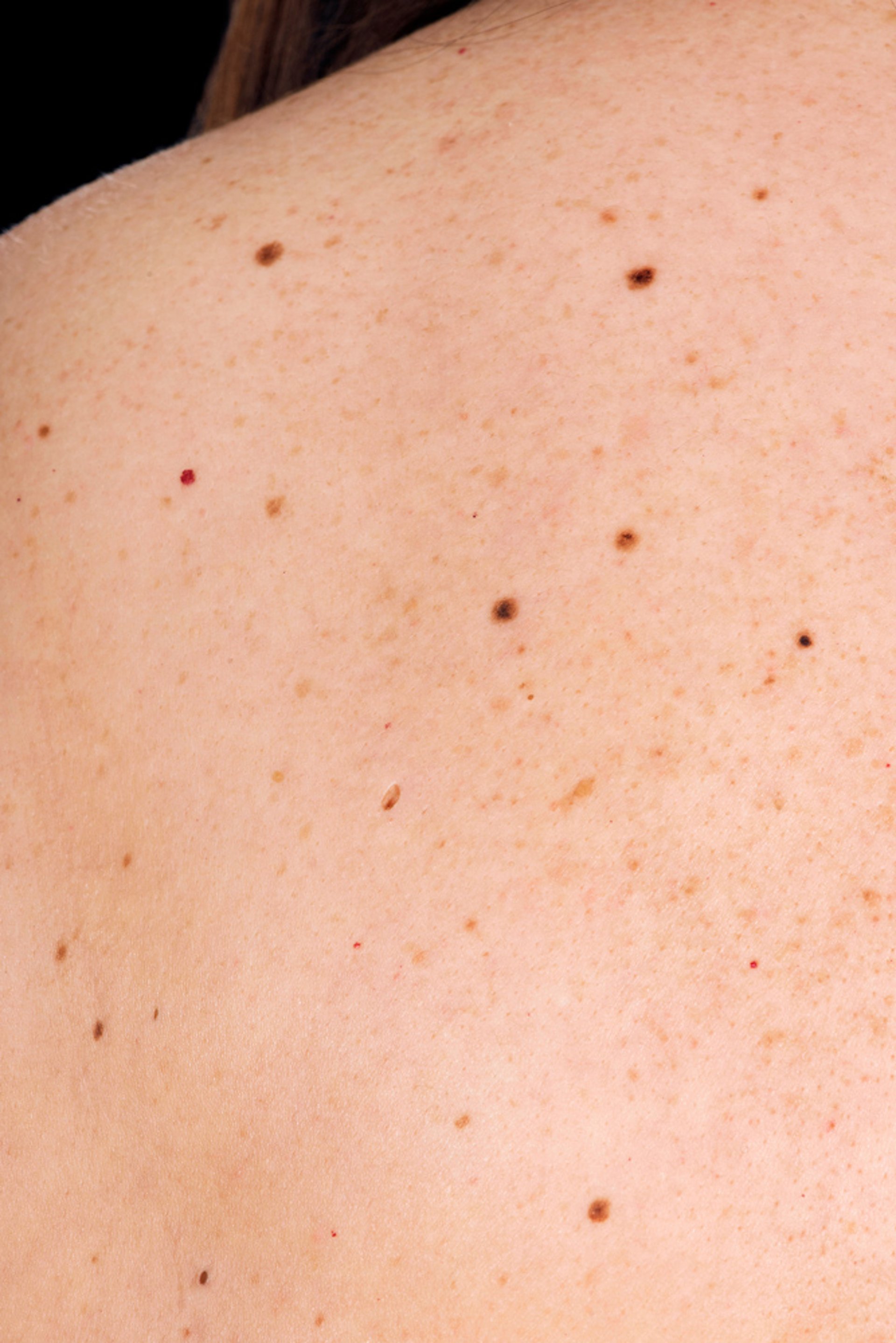
Polarized light and immersion contact dermoscopy may be useful for distinguishing melanomas from benign lesions. However, biopsy and histologic evaluation should be considered if moles have certain characteristics of concern (known as the ABCDEs of melanoma):
A: Asymmetry—asymmetric appearance
B: Borders—irregular borders (ie, not round or oval)
C: Color—color variation within the mole, unusual colors, or a color significantly different or darker than the patient's other moles
D: Diameter—> 6 mm
E: Evolution—a new mole in a patient > 30 years of age or a changing mole
Patients at risk can be taught self-examination to detect changes in existing moles and to recognize features suggesting melanoma.
Other red flags include
Recent enlargement or change in shape
Change in surface characteristics or consistency
Signs of inflammation in surrounding skin, with possible bleeding, ulceration, itching, or tenderness
However, recent enlargement, darkening, ulceration, or bleeding usually indicates that the melanoma has already invaded the skin deeply.
If doubt exists, biopsy should include the full depth of the dermis and extend slightly beyond the edges of the lesion. Because earlier diagnosis can be lifesaving and features of melanoma can be variable, even slightly suspect lesions should be biopsied. Earlier diagnosis of melanoma is possible if biopsy specimens can be obtained from lesions having variegated colors (eg, brown or black with shades of red, gray, or blue), irregular elevations that are visible or palpable, and borders with angular indentations or notches.
Biopsy should be excisional for most lesions except those on anatomically sensitive or cosmetically important areas; in these cases, a broad shave biopsy can be done. For broader lesions such as lentigo maligna, representative shave biopsies from several areas can increase the diagnostic yield. By doing step sections, the pathologist can determine the maximal thickness of the melanoma. Definitive radical surgery should not precede histologic diagnosis.
BRAF inhibitor, for metastatic melanomas bearing a V600 mutation in the BRAF gene.
Differential diagnosis includes basal cell carcinomas and squamous cell carcinomas, seborrheic keratoses, atypical moles, blue nevi, dermatofibromas, moles, hematomas (especially on the hands or feet), venous lakes, pyogenic granulomas, and warts with focal thromboses.
Staging
The staging of melanoma is based on clinical and pathologic criteria and closely corresponds to the traditional tumor-node-metastasis (TNM) classification system. The staging system classifies melanomas based on local, regional, or distant disease:
Stages I and II: Localized primary melanoma
Stage III: Metastasis to regional lymph nodes
Stage IV: Distant metastatic disease
Stage strongly correlates with survival. Sentinel lymph node biopsy (SLNB), a minimally invasive microstaging technique, is a major advance in the ability to stage cancers more accurately. Recommended staging studies depend on the Breslow depth (how deeply tumor cells have invaded) and histologic characteristics of the melanoma; ulceration indicates higher risk in melanomas that are < 0.8 mm Breslow depth (see table Staging of Melanoma Based on Thickness and Ulceration). Staging studies may include sentinel lymph node biopsy, laboratory tests (eg, complete blood count, lactate dehydrogenase, liver tests), chest x-ray, CT, and positron emission tomography (PET) and are done by a coordinated team that includes dermatologists, oncologists, general surgeons, plastic surgeons, and dermatopathologists.
Staging of Localized Melanoma Based on Thickness and Ulceration
Stage | Thickness | Ulceration Status |
|---|---|---|
0 | Intraepithelial or in situ melanoma | Not applicable |
IA | < 0.8 mm | Without ulceration |
IB | < 0.8 mm | With ulceration |
0.8–1.0 mm | With or without ulceration | |
IIA | > 1.0–2.0 mm | Without ulceration |
IIB | > 1.0–2.0 mm | With ulceration |
IIIA | > 2.0–4.0 mm | Without ulceration |
IIIB | > 2.0–4.0 mm | With ulceration |
IVA | > 4.0 mm | Without ulceration |
IVB | > 4.0 mm | With ulceration |
Data from Keung EZ, Gershenwald JE: The eighth edition American Joint Committee on Cancer (AJCC) melanoma staging system: implications for melanoma treatment and care. Expert Rev Anticancer Ther 18(8):775-784, 2018. doi: 10.1080/14737140.2018.1489246 | ||
Treatment of Melanoma
Surgical excision
(See also the American Academy of Dermatology Association’s 2019 guidelines of care for the management of primary cutaneous melanoma.)
Treatment of melanoma is primarily by surgical excision (wide local excision). Although the width of margins is debated, most experts agree that a 1-cm lateral tumor-free margin is adequate for lesions < 0.8 mm thick. In tumors < 0.8 mm thick but with ulceration, sentinel lymph node biopsy can be considered. Thicker lesions may deserve larger margins, more radical surgery, and sentinel lymph node biopsy.
Lentigo maligna melanoma and lentigo maligna are usually treated with wide local excision and, if necessary, skin grafting. Intensive radiation therapy is much less effective.
Spreading or nodular melanomas have usually been treated with wide local excision. Lymph node dissection is recommended when nodes are involved clinically or on histologic evaluation of sentinel lymph node biopsy.
Metastatic disease
Treatment of metastatic melanoma typically includes
Immunotherapy
Molecular targeted therapy
Radiation therapy
Rarely surgical resection
All of these treatments should be considered for all patients who have metastatic melanoma. Final decisions are generally individualized by an oncologist and may depend on availability.
Metastatic disease is generally inoperable, but in certain cases, localized and regional metastases can be excised to help eliminate residual disease and prolong survival.
Molecular targeted therapy1, 2, 3).
Cytotoxic chemotherapy has not been shown to improve survival in patients with metastatic disease and is normally reserved for patients who do not have other options.
Radiation therapy may be used when positive resection margins are not possible because of the location, in desmoplastic melanoma, in locally recurrent melanoma after re-excision, and in palliate brain metastases, but the response is poor (4, 5).
The following are under study:
Infusion of lymphokine-activated killer cells or antibodies (for advanced-stage disease)
Treatment references
1. Long GV, Flaherty KT, Stroyakovskiy D, et al: Dabrafenib plus trametinib versus dabrafenib monotherapy in patients with metastatic BRAF V600E/K-mutant melanoma: Long-term survival and safety analysis of a phase 3 study. Ann Oncol 28(7):1631–1639, 2017. doi: 10.1093/annonc/mdx176. Clarification and additional information. Ann Oncol 30(11):1848, 2019.
2. Long GV, Stroyakovskiy D, Gogas H, et al: Dabrafenib and trametinib versus dabrafenib and placebo for Val600 BRAF-mutant melanoma: A multicentre, double-blind, phase 3 randomised controlled trial. Lancet 386(9992):444–451, 2015. doi: 10.1016/S0140-6736(15)60898-4
3. Long GV, Stroyakovskiy D, Gogas H, et al: Combined BRAF and MEK inhibition versus BRAF inhibition alone in melanoma. N Engl J Med 371(20):1877–1888, 2014. doi: 10.1056/NEJMoa1406037
4. Mendenhall WM, Shaw C, Amdur RJ, et al: Surgery and adjuvant radiotherapy for cutaneous melanoma considered high-risk for local-regional recurrence. Am J Otolaryngol 34(4):320–322, 2013. doi: 10.1016/j.amjoto.2012.12.014
5. Rule WG, Allred JB, Pockaj BA, et al: Results of NCCTG N0275 (Alliance): A phase II trial evaluating resection followed by adjuvant radiation therapy for patients with desmoplastic melanoma. Cancer Med 5(8):1890–1896, 2016. doi: 10.1002/cam4.783
Prognosis for Melanoma
Melanoma may spread rapidly, causing death within months of its recognition, yet the 5-year cure rate of early, very superficial lesions is very high. Thus, cure depends on early diagnosis and early treatment. The 5-year survival rates range from 99.6% for localized melanomas to 73.9% with regional spread and 35.1% with distant metastases (1).
For tumors of cutaneous origin (not central nervous system and subungual melanomas) that have not metastasized, the survival rate varies depending on the thickness of the tumor at the time of diagnosis.
Mucosal melanomas (especially anorectal melanomas) have a poor prognosis, although they often seem quite limited when discovered. They account for a higher proportion of melanomas in Black, Hispanic, and Asian people compared with White people.
Degree of lymphocytic infiltration, which represents reaction by the patient’s immunologic defense system, may correlate with the level of invasion and prognosis. Chances of cure are maximal when lymphocytic infiltration is limited to the most superficial lesions and decrease with deeper levels of tumor cell invasion, ulceration, and vascular or lymphatic invasion.
A commercially available test of gene expression can help determine whether patients who have stage I or II melanomas are at high or low risk of metastases. This test has not yet been added to consensus guidelines; using it to determine whether patients should receive immunotherapy is not recommended at this time.
Prognosis reference
1. National Institutes of Health: Cancer Stat Facts: Melanoma of the Skin. Accessed October 10, 2023.
Prevention of Melanoma
Because melanoma is associated with ultraviolet (UV) exposure, a number of measures are recommended to limit exposure.
Sun avoidance: Seeking shade, minimizing outdoor activities between 10 AM and 4 PM (when sun's rays are strongest), and avoiding sunbathing and the use of tanning beds
Use of protective clothing: Long-sleeved shirts, pants, and broad-brimmed hats
Use of sunscreen: At least sun protection factor (SPF) 30 with broad-spectrum UVA/UVB protection, used as directed (ie, reapplied every 2 hours and after swimming or sweating); should not be used to prolong sun exposure
However, current evidence is inadequate to determine whether these measures reduce incidence or mortality of melanoma.
Key Points
Melanoma accounts for < 2% of total skin cancers diagnosed in the United States but causes most skin cancer deaths.
Melanoma can develop in the skin, mucosa, conjunctiva, choroid layer of the eye, leptomeninges, and nail beds.
Although melanoma can develop from a typical or atypical mole, most do not.
Physicians (and patients) should monitor moles for changes in size, shape, borders, color, or surface characteristics and for bleeding, ulceration, itching, and tenderness.
Biopsy even slightly suspect lesions.
Excise melanomas whenever feasible, particularly when melanomas have not metastasized.
More Information
The following English-language resource may be useful. Please note that THE MANUAL is not responsible for the content of this resource.
American Cancer Society: Cancer Facts & Figures 2023


