


Cutaneous larva migrans is the skin manifestation of hookworm infestation. Diagnosis is clinical. Treatment is oral or topical antihelminthic therapy.
Cutaneous larva migrans is caused by Ancylostoma species, most commonly dog or cat hookworm Ancylostoma braziliense.
Hookworm ova in dog or cat feces develop into infective larvae when left in warm moist ground or sand. Transmission occurs when skin directly contacts contaminated soil or sand and larvae penetrate unprotected skin, usually of the feet, legs, buttocks, or back.
Cutaneous larva migrans occurs worldwide but most commonly in tropical environments. Emergence of this condition in previously naive countries is thought to be due to climate change (1).
© Springer Science+Business Media
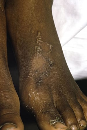
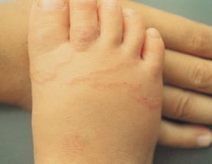
Image courtesy of Karen McKoy, MD.


© Springer Science+Business Media


Image courtesy of Karen McKoy, MD.
Cutaneous larva migrans causes intense pruritus. Signs are erythema and papules at the site of entry, followed by a winding, threadlike subcutaneous trail of reddish brown inflammation. Patients may also develop papules and vesicles resembling folliculitis, called hookworm folliculitis.
Cutaneous larva migrans may be complicated by a self-limiting pulmonary reaction called Löffler syndrome (patchy pulmonary infiltrates and peripheral blood eosinophilia) (2).
Diagnosis of cutaneous larva migrans is by history and clinical appearance.
General references
1. Ahmed A, Hemaida MA, Hagelnur AA, et al: Sudden emergence and spread of cutaneous larva migrans in Sudan: A case series calls for urgent actions. IDCases 32:e01789, 2023. doi: 10.1016/j.idcr.2023.e01789
2. Podder I, Chandra S, Gharami RC: Loeffler's syndrome following cutaneous larva migrans: An uncommon sequel. Indian J Dermatol 61(2):190–192, 2016. doi: 10.4103/0019-5154.177753
Treatment of Cutaneous Larva Migrans
Oral or topical antihelminthic therapy
Although the infection usually resolves spontaneously after a few weeks, discomfort and the risk of secondary bacterial infection warrant treatment.
albendazole 10% ointment (compounded) and thiabendazole 15% liquid or cream (compounded) can be used as alternatives.


