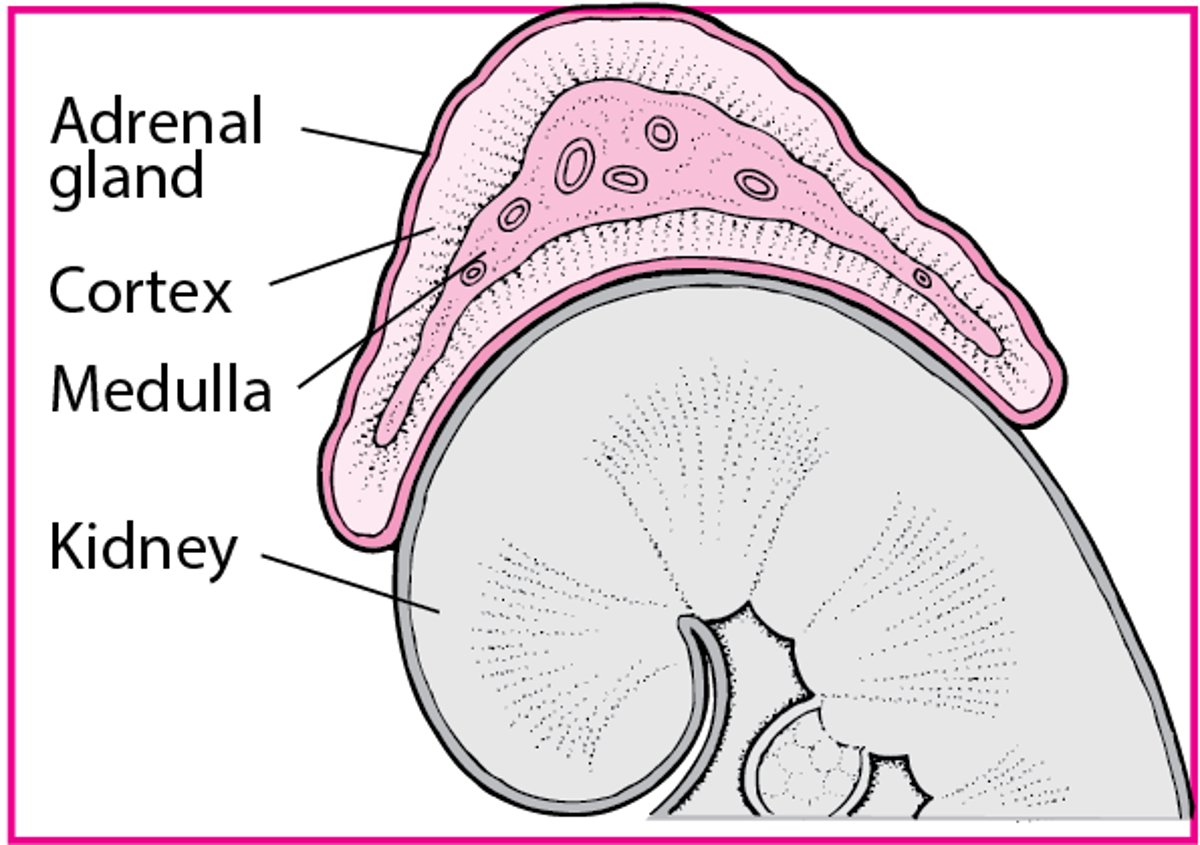
 |
The immune system distinguishes self from nonself and eliminates potentially harmful nonself molecules and cells from the body. The immune system also has the capacity to recognize and destroy abnormal cells that derive from host tissues (see Tumor Immunology: Host Response to Tumors). Any molecule capable of being recognized by the immune system is considered an antigen (Ag).
Anatomic barriers, part of the body's external defenses, must be overcome before the immune system is activated to destroy foreign antigens. These barriers include the outer, keratinized epidermis; mucus of the respiratory, GI, and urogenital tracts; and cilia on respiratory epithelial cells, which help expel particles and organisms trapped by mucus. Each has specialized immune functions. For example, in the skin, keratinocytes secrete antimicrobial peptides (defensins), and sebaceous and sweat glands secrete microbe-inhibiting substances (eg, lactic acid, fatty acids). Also, many immune cells (eg, mast cells, intraepithelial lymphocytes, Ag-sampling Langerhans' cells) reside in the skin. Mucus in respiratory, GI, and GU tracts contains antimicrobial substances, such as lysozyme, lactoferrin, and secretory IgA antibody (SIgA). Breaching of anatomic barriers can trigger 2 types of immune response: innate and acquired.
Innate (natural or nonspecific) immunity does not require prior exposure to an Ag (ie, memory) to be effective. Components include phagocytic cells (neutrophils and monocytes in the blood, macrophages and dendritic cells in tissues) that ingest and destroy invading Ags; Ag-presenting cells (macrophages, dendritic cells) that present fragments of ingested Ags to T cells, which are part of acquired immunity; natural killer cells that kill virus-infected cells and some tumor cells; and certain polymorphonuclear leukocytes (eosinophils, basophils, mast cells) that release inflammatory mediators.
Acquired (adaptive or specific) immunity remembers past exposures and is Ag-specific. Components include antibody (Ab) and T and B cells (lymphocytes). Acquired immunity derived from certain T-cell responses is called cellular immunity; immunity derived from B-cell responses is called humoral immunity because B cells secrete soluble Ag-specific Ab.
Innate and acquired immune responses interact; eg, IgE (acquired immunity) binds to mast cells or basophils (innate immunity) to produce an allergic response. Many molecular components participate in innate and acquired immunity; they include complement, cytokines, and acute phase reactants.
Successful immune defense requires activation, regulation, and resolution of the immune response.
Activation:
The immune system is activated when a foreign Ag is recognized by circulating Abs or cell surface receptors. These receptors may be highly specific (eg, Ig expressed on B cells or bound to phagocytes) or broadly specific (eg, pattern-recognition receptors on dendritic cells, including mannose, scavenger, and Toll-like receptors, which recognize common microbial pathogen-associated molecular patterns in ligands such as gram-negative lipopolysaccharide, unmethylated cytosine-guanosine dinucleotides [CpG motifs], and peptidoglycans). Ab-Ag and complement-microorganism complexes can also bind to surface receptors for the crystallizable fragment (Fc) region of IgG (FcγR) and for C3b and iC3b.
Once recognized, an Ag, Ag-Ab complex, or complement-microorganism complex is phagocytosed. Some microorganisms are killed after they are phagocytosed; others (eg, mycobacteria) inhibit the phagocyte's ability to kill them once they are engulfed. In such cases, T cell–derived cytokines, particularly interferon-γ (IFN-γ), stimulate the phagocyte to produce lytic enzymes and other microbicidal macrophage products, which kill the microorganism.
Unless Ag is rapidly phagocytosed and entirely degraded (an uncommon event), the acquired immune response is recruited. This response begins in the spleen for circulating Ag, in regional lymph nodes for tissue Ag, and in mucosa-associated lymphoid tissues (eg, tonsils, adenoids, Peyer's patches) for mucosal Ag. For example, Langerhans' dendritic cells in the skin phagocytose Ag and migrate to local lymph nodes; there, peptides derived from the Ag are expressed on the cell surface within class II major histocompatibility complex (MHC) molecules, which present the peptide to CD4 helper T (TH) cells. When the TH cell engages the complex, the cell expresses receptors for the cytokine IL-2 and secretes several cytokines. A subset of TH cells (TH1) secretes IFN-γ, IL-2, and lymphotoxin, which facilitate macrophage and cytotoxic T-cell responses; another subset, TH2 cells, secretes IL-4, IL-5, IL-6, IL-10, and IL-13, which stimulate antibody production by B-cells.
In contrast to class II MHC molecules, which present extracellular Ag to CD4 TH cells, class I MHC molecules present intracellular Ag (eg, viruses) to CD8 cytotoxic T cells. The activated cytotoxic T cell then kills the infected cell.
Regulation:
The immune response must be regulated to prevent overwhelming damage to the host (eg, anaphylaxis, widespread tissue destruction). Regulatory T cells help control the immune response via secretion of immunosuppressive cytokines, such as IL-10 and transforming growth factor-β (TGF-β), or via a poorly defined cell contact mechanism. These regulatory cells help prevent autoimmune responses and probably help resolve ongoing responses to nonself Ag.
Resolution:
The immune response resolves when Ag is sequestered and eliminated from the body. Without stimulation by Ag, cytokine secretion ceases, and activated cytotoxic T cells undergo apoptosis. Apoptosis tags a cell for immediate phagocytosis, which prevents spillage of the cellular contents and development of subsequent inflammation. T and B cells that have differentiated into memory cells are spared this fate.
Last full review/revision November 2005
Content last modified November 2005
|  |