Patients & CaregiversHealthcare ProfessionalsWorldwide
HomeAbout MerckProductsNewsroomInvestor RelationsCareersResearchLicensingThe Merck Manuals
THE MERCK MANUAL MEDICAL LIBRARY: The Merck Manual of Diagnosis and Therapy
Tips for better results
ABCDEFGHI
JKLMNOPQR
STUVWXYZ

Section

Subject

Topics

Introduction

Update Me

(See also Hearing Loss; see also Nose and Paranasal Sinus Disorders; see also Optic Nerve Disorders; and see also Approach to the Neurologic Patient). (See also Autonomic Nervous System: Horner's Syndrome.)

Dysfunction of certain cranial nerves may affect the eye, pupil, optic nerve, or extraocular muscles and their nerves; thus, they can be considered cranial nerve disorders, neuro-ophthalmologic disorders, or both. Neuro-ophthalmologic disorders may also involve dysfunction of the central pathways that control and integrate ocular movement and vision. 
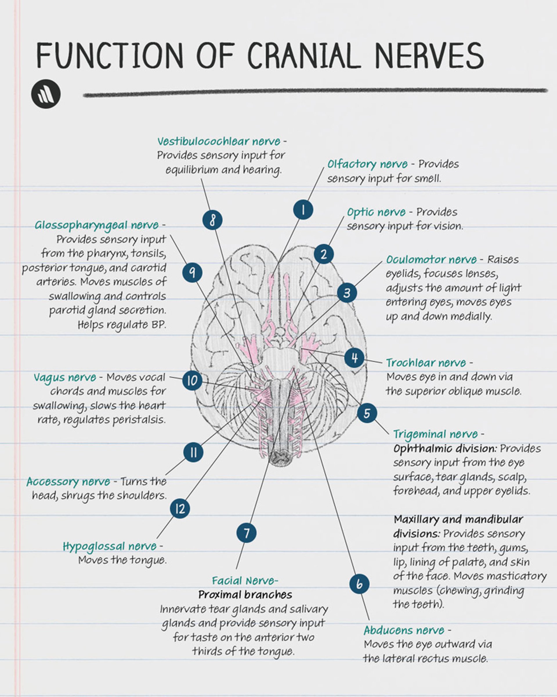
Cranial nerve disorders can also involve dysfunction of smell, vision, chewing, facial sensation or expression, taste, hearing, balance, swallowing, phonation, head turning and shoulder elevation, or tongue movements (see Table 1: Neuro-ophthalmologic and Cranial Nerve Disorders: Cranial NervesTables). One or more cranial nerves may be affected.

Table 1

PDF Cranial Nerves

This table is presented as a PDF and requires the free Adobe PDF reader. Get Adobe Reader

Causes and symptoms of neuro-ophthalmologic and cranial nerve disorders overlap. Both types of disorders can result from tumors, inflammation, trauma, systemic disorders, and degenerative or other processes, causing such symptoms as vision loss, diplopia, ptosis, pupillary abnormalities, periocular pain, facial pain, or headache.

Diagnosis and Treatment

Evaluation includes the following:

Visual system examination includes ophthalmoscopy and testing of visual acuity, visual fields (see Table 3: Approach to the Ophthalmologic Patient: Causes of TearingTables), pupils (see Table 2: Neuro-ophthalmologic and Cranial Nerve Disorders: Common Pupillary AbnormalitiesTables), and eye movements (ocular motility—see Table 3: Neuro-ophthalmologic and Cranial Nerve Disorders: Common Disturbances of Ocular MotilityTables). As part of this testing, the 2nd, 3rd, 4th, and 6th cranial nerves are examined (see also Approach to the Neurologic Patient: Cranial nerves). Neuroimaging with CT or MRI is also usually required.

The following parts of the visual examination are of particular interest in diagnosing neuro-ophthalmologic and cranial nerve disorders:

Pupils are inspected for size, equality, and regularity. Normally, the pupils constrict promptly (within 1 sec) and equally during accommodation and during exposure to direct light and to light directed at the other pupil (consensual light reflex). Testing pupillary response to consensual light via a swinging flashlight test can determine whether a defect is present. Normally, the degree of pupillary constriction does not change as the flashlight is swung from eye to eye.

  • If a relative afferent defect (deafferented pupil, afferent pupillary defect, or Marcus Gunn pupil) is present, the pupil paradoxically dilates when the flashlight swings to the side of the defect. A deafferented pupil constricts in response to consensual but not to direct light.
  • If an efferent defect is present, the pupil responds sluggishly or does not respond to both direct and consensual light.

Eye movements are checked by having the patient hold the head steady, while tracking the examiner's finger as it moves to the far right, left, upward, downward, diagonally to either side, and inward toward the patient's nose (to assess accommodation). However, such examination may miss mild paresis of ocular movement sufficient to cause diplopia.

Diplopia may indicate a defect in bilateral coordination of eye movements (eg, in neural pathways) or in the 3rd (oculomotor), 4th (trochlear), or 6th (abducens) cranial nerve. If diplopia persists when one eye is closed (monocular diplopia), the cause is probably a nonneurologic eye disorder (see Approach to the Ophthalmologic Patient: Diplopia). If diplopia disappears when either eye is closed (binocular diplopia), the cause is probably a disorder of ocular motility. The 2 images are furthest apart when the patient looks in the direction served by the paretic eye muscle (eg, to the left when the left lateral rectus muscle is paretic). The eye that, when closed, eliminates the more peripheral image is paretic. Placing a red glass over one eye can help identify the paretic eye. When the red glass covers the paretic eye, the more peripheral image is red (see also Approach to the Ophthalmologic Patient: Diplopia).

Treatment of neuro-ophthalmologic and cranial disorders depends on the cause.

Table 2

Common Pupillary Abnormalities

Finding

Explanation

Asymmetry of 1–2 mm between pupils, preserved light responses, and no symptoms (physiologic anisocoria)

Normal variant

Asymmetry, impaired light responses, and preserved response to accommodation (light-near dissociation or Argyll Robertson pupil)

Neurosyphilis (possibly)

Bilateral constriction

Opioids

Organophosphate or cholinergic toxins

Pontine hemorrhage

Miotic eye drops for glaucoma (most common; causing unilateral constriction if single eye is dosed)

Bilateral dilation with preserved light reflexes

Hyperadrenergic states (eg, withdrawal syndromes, drugs such as sympathomimetics or cocaine, thyrotoxicosis)

Bilateral dilation with impaired light response

Mydriatic eye drops such as sympathomimetics (eg, phenylephrine Some Trade Names
NEO-SYNEPHRINE
Click for Drug Monograph
) and cycloplegics (eg, cyclopentolate Some Trade Names
AK-PENTOLATE
CYCLOGYL
CYLATE
Click for Drug Monograph
, tropicamide Some Trade Names
MYDRAL
MYDRIACYL
TROPICACYL
Click for Drug Monograph
, homatropine Some Trade Names
ISOPTO
Click for Drug Monograph
, atropine Some Trade Names
ATROPEN
ATROPINE-CARE
SAL-TROPINE
Click for Drug Monograph
)

Brain herniation

Hypoxic or ischemic encephalopathy

Unilateral dilation with afferent pupillary defect

Lesions of the eye, retina, or 2nd cranial (optic) nerve

Unilateral dilation with efferent pupillary defect

Third cranial (oculomotor) nerve palsies, often due to compression (eg, due to aneurysm of the posterior communicating artery or to transtentorial herniation)

Iris trauma (also irregular pupil)

Mydriatic eye drops*

Unilateral dilation with minimal or slow direct and consensual light reflexes and pupil constriction in response to accommodation

Tonic (Adie's) pupil†

*Transtentorial herniation and use of mydriatic eye drops can often be distinguished by instilling a drop of pilocarpine Some Trade Names
ISOPTO CARPINE
PILOPINE HS
SALAGEN
Click for Drug Monograph
ocular solution into the dilated pupil; no constriction in response suggests mydriatic eye drops.

†Tonic pupil is permanent but nonprogressive abnormal dilation of the pupil due to damage of the ciliary ganglion. It typically occurs in women aged 20 to 40. Onset is usually sudden. The only findings are slight blurring of vision, impaired dark adaptation, and sometimes absent deep tendon reflexes.

Table 3

Common Disturbances of Ocular Motility

Clinical Finding

Syndrome

Common Causes

Pareses

Paresis of horizontal gaze in one direction

Conjugate horizontal gaze palsy

Lesion in the ipsilateral pontine horizontal gaze center or in the contralateral frontal cortex

Paresis of horizontal gaze in both directions

Complete (bilateral) horizontal gaze palsy

Wernicke's encephalopathy

Large bilateral pontine lesion affecting both horizontal gaze centers

Bilateral paresis of all horizontal eye movements except for abduction of the eye contralateral to the lesion; convergence unaffected

One-and-a-half syndrome

Lesion in the medial longitudinal fasciculus and ipsilateral pontine horizontal gaze center

Unilateral or bilateral paresis of eye adduction in horizontal lateral gaze but not in convergence

Internuclear ophthalmoplegia

Lesion in the medial longitudinal fasciculus

Bilateral paresis of upward eye movement with dilated pupils, loss of the pupillary light response despite preservation of pupillary accommodation and constriction with convergence, downward gaze preference, and downbeating nystagmus

Parinaud's syndrome (a type of conjugate vertical gaze palsy)

Pineal tumor

Midbrain infarct

Bilateral paresis of downward eye movements

Conjugate downward gaze palsy

Progressive supranuclear palsy

Unilateral eye deviation (resting position is down and out); unilateral paresis of eye adduction, elevation, and depression; ptosis; and often a dilated pupil

3rd cranial nerve palsy

Aneurysms

Oculomotor nerve or midbrain ischemia

Trauma

Transtentorial herniation

Unilateral paresis of downward and inward (nasal) eye movement, which may be subtle, causing symptoms (difficulty looking down and inward)

Head tilt sign (patient tilts head opposite the side of the affected eye)

4th cranial nerve palsy

Idiopathic

Head trauma

Ischemia

Congenital

Unilateral paresis of eye abduction

6th cranial nerve palsy

Idiopathic

Infarct

Vasculitis

Increased intracranial pressure

Wernicke's encephalopathy

Multiple sclerosis

Skew deviation (vertical misalignment of the eyes)

Partial and unequal involvement of 3rd cranial nerve nuclei, vertical gaze center, or median longitudinal fasciculus

Brain stem lesion anywhere from midbrain to medulla

Weakness or restriction of all extraocular muscles

External ophthalmoplegia

Dysfunction of eye muscles or of neuromuscular junction

Usual causes include the following:

  • Myasthenia gravis
  • Graves disease
  • Botulism
  • Mitochondrial myopathies (eg, Kearn-Sayre syndrome)
  • Oculopharyngeal dystrophy
  • Myotonic dystrophy
  • Orbital pseudotumor

Involuntary or abnormal movements

Rythmic involuntary movements, usually bilateral

Nystagmus

see Sidebar 1: Approach to the Patient With Ear Problems: NystagmusSidebars

Fast downward jerk and slow upward return to midposition

Ocular bobbing

Extensive pontine destruction or dysfunction

Gaze overshoot followed by several oscillations

Ocular dysmetria

Cerebellar pathway disorders

Burst of rapid horizontal oscillations about a point of fixation

Ocular flutter

Many causes:

  • Postanoxic encephalopathy
  • Occult neuroblastoma
  • Paraneoplastic effects
  • Ataxia-telangiectasia
  • Viral encephalitis
  • Toxic effects of drugs

Rapid, conjugate, multidirectional, chaotic movements, often with widespread myoclonus

Opsoclonus

Many causes (same as for ocular flutter, above)

Last full review/revision October 2007 by Michael Jacewicz, MD

Content last modified October 2007

Back to Top

Next: Bell's Palsy

Audio
Figures
Photographs
Tables
Videos
Contact UsSite MapPrivacy PolicyTerms of UseCopyright 1995-2007 Merck & Co., Inc.