


Common skin disorders and infections can cause cutaneous penile lesions (see table Causes of Cutaneous Penile Lesions).
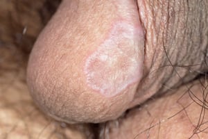
Balanitis xerotica obliterans
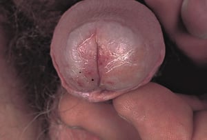
This lesion, another name for lichen sclerosus et atrophicus in men, is an indurated, blanched area near the tip of the glans surrounding and often constricting the meatus. It results from chronic inflammation and may lead to phimosis, paraphimosis, or urethral stricture
© Springer Science+Business Media
© Springer Science+Business Media
© Springer Science+Business Media
© Springer Science+Business Media
DR P. MARAZZI/SCIENCE PHOTO LIBRARY
DR P. MARAZZI/SCIENCE PHOTO LIBRARY


© Springer Science+Business Media


© Springer Science+Business Media


© Springer Science+Business Media


© Springer Science+Business Media


DR P. MARAZZI/SCIENCE PHOTO LIBRARY


DR P. MARAZZI/SCIENCE PHOTO LIBRARY
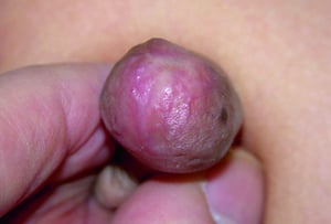
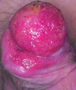
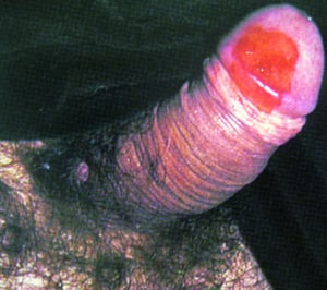
Carcinoma in situ
Carcinoma in situ can include
Erythroplasia of Queyrat: Squamous cell carcinoma in situ of the glans or prepuce
Bowen disease of the penis: Squamous cell carcinoma in situ of the penile skin
Bowenoid papulosis: Associated with human papillomavirus (particularly HPV types 16 and 18)
Erythroplasia of Queyrat and Bowen disease of the penis are well-circumscribed areas of reddish, velvety pigmentation in the genital area, usually on the glans or at the corona, primarily in uncircumcised men.
Paget disease of the nipple (not to be confused with Paget disease of bone) is a rare intraepithelial adenocarcinoma that can occur in extramammary locations, including the penis.
Bowenoid papulosis involves smaller, often multiple papules on the shaft of the penis.
These conditions are considered intraepithelial neoplasia or carcinoma in situ and should be biopsied.
Penile lichen planus
This lesion occurs as small plaques, papules or macules, sometimes annular, on the glans or shaft and may be mistaken for pemphigoid or erythema multiforme. Pruritus is common.
Penogingival syndrome in men (and vulvovaginal gingival syndrome in women) is a more severe form of erosive lichen planus. It occurs on both oral and genital mucosa. Ulcers may develop and cause pain.
Lichen planus usually resolves spontaneously. If asymptomatic, it may not require treatment. Topical corticosteroids may help relieve symptoms.
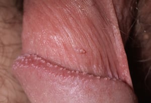
Pearly penile papules
These papules are small, harmless angiofibromas that appear on the corona of the penis as dome-shaped or hairlike projections and tend to be skin-colored. They may also appear on the distal shaft. They are common, occurring in up to 10% of men. They are not associated with human papillomavirus, although they may be mistaken for genital warts. Treatment is not required.
Contact dermatitis of the penis
Contact dermatitis of the penis has become more common with the widespread use of latex condoms. Dermatitis appears as red, pruritic lesions, sometimes with weeping or fissures. Treatment is with topical corticosteroids and use of nonlatex condoms (but not natural condoms, which do not provide adequate protection against HIV). Mild over-the-counter corticosteroids can be tried first, with use of middle- or high-potency prescription preparations as needed.


